CONTATO & EQUIPE
Para mais informações sobre a linha de luz, entre em contato.


The XRF beamline is an experimental station dedicated to X-ray Fluorescence Microscopy (XRFM), X-ray Fluorescence Tomography (XFCT) and Total Reflection X-ray Fluorescence (TXRF) analysis in the hard X-rays energy range (5 to 20 keV). The beamline’s focus is on the determination and mapping of trace chemical elements in samples with applications in the fields of analytical chemistry, biomedicine, environmental geochemistry and materials science.
XRF’s source is a 1.67T bending magnet. The monochromator vacuum chamber can be laterally displaced, so that the whole synchrotron spectra can also be used to excite the samples. The experimental facilities include one station consisting of a high vacuum chamber in which grazing incidence x-ray fluorescence experiments can be carried out. The chamber is equipped with remote-controlled XY$\rm \theta$y sample stages and a HPGe solid state detector, optimised for the detection of light element. The whole setup is mounted in the motorised lift table, which allows vertical positioning of the instruments on the plane where the incoming beam is mostly linearly polarized.
Applications include 2D XRF mapping and speciation of trace elements at 20 microns resolution, 3D information of elements in volumetric samples, analysis of very small masses deposited on flat substrate, trace impurities on surfaces of flat samples, chemical depth profiling surface analysis (from sub nm to mm range).
Para mais informações sobre a linha de luz, entre em contato.
As técnicas e configurações experimentais a seguir estão disponíveis nesta linha de luz. Para saber mais sobre as limitações e requerimentos das técnicas, contate o coordenador da linha de luz antes de submeter sua proposta.
X-ray Fluorescence (XRF) is a well-established bulk analytical method for qualitative as well as quantitative determination of several elements in a sample, independent of their chemical form. Depending on the detection system configuration, can be classified as Energy Dispersive (EDXRF) or Wavelength Dispersive (WDXRF) systems.
Setup: X-ray Fluorescence Microscopy (XRFM)
This setup allows performing experiments with an X-ray microbeam. The microfocusing optics consists of a pair of bendable mirrors arranged in the so called Kirkpatrick-Baez configuration (KB). The X-ray microbeam is around 12 microns (vertical) by 22 microns (horizontal) in size. Fully remote-controlled XYZz sample stages can operate in air or under a nitrogen gas environment. The $\rm \mu$-XRF setup also includes an optical microscope and a Silicon Drift Detector (SSD).
Other setups include:
| Elemento | Tipo | Posição [m] | Descrição |
|---|---|---|---|
| SOURCE | Bending Magnet | 0.0 | Bending Magnet D09 exit B (15°), 1.67 T , 0.92 mm x 0.57 mm |
| Mono | Double Crystal Monochromator | 11.2 | Si(111), Si(220), channel-cut type |
| M1 | Elliptical Vertical Micro- focusing Mirror | 14.9 | Rh coated, RT=334m (center), θ=4 mrad |
| M2 | Elliptical Horizontal Microfocusing Mirror | 15.3 | Rh coated, RT = 176m (center), θ=4 mrad |
| Parâmetro | Valor | Condição |
|---|---|---|
| Energy range [keV] | 5-20 | Si(111) / Si(220) |
| Energy resolution [ΔE/E] 10-4 | 10-4 | Si(111) |
| Beam size at sample [µm2, FWHM] | 22 x 12 | at 10 keV |
| Beam divergence at sample [mrad2, FWHM] | 10 x 1 | at 10 keV |
| Flux density at sample [ph/s/mm2/100 mA] | 2 x 10⁸ | at 10 keV |
| Flux at focal spot [ph/s/100mA] | 2 x 10¹² | White beam |
| Instrumento | Tipo | Modelo | Fabricante | Especificações |
|---|---|---|---|---|
| Detector | Silicon drift | AXAS-A | 30 mm2 SDD, FWHM ≤ 139eV at 5.9keV | KETEK GmbH |
| Detector | Silicon drift | AXAS-A | 7 mm2 SDD, FWHM ≤ 139eV at 5.9keV | KETEK GmbH |
| Detector | HPGe | GUL0035 | 30 mm2 Ultra-LGe, FWHM = 140 eV at 5.9keV | Canberra |
| Cryostat | Cryostream Cooler | 700 Series | Minimum of 173K, with a gas stability of 0.1K | Oxford |
All beamline controls are done through EPICS (Experimental Physics and Industrial Control System), running on a PXI from National Instruments. The data acquisition is done using a Red Hat workstation with the Py4Syn, developed at LNLS by SOL group. CSS (Control System Studio) are used as a graphical interface to display and control the beamline devices. Point-to-point or continuous scanning (“on-the-fly”) modes of operation can be used for data acquisition in 2D/3D experiments.
Usuários devem declarar a utilização das instalações do LNLS em qualquer publicação, como artigos, apresentações em conferências, tese ou qualquer outro material publicado que utilize dados obtidos na realização de sua proposta.
PÉREZ, C. A., RADTKE, M., SÁNCHEZ, H. J., TOLENTINO, H., NEUENSCHWANDER, R. T., BARG, W., RUBIO, M., BUENO, M. I. S., RAIMUNDO I. M. & ROHWEDDER, J. J. R., Synchrotron Radiation X-Ray Fluorescence at the LNLS: Beamline Instrumentation and Experiments, X-Ray Spectrometry 28, 320–326 (1999). doi: 10.1002/(SICI)1097-4539(199909/10)28:53.0.CO;2-1
The x-ray fluorescence beamline of the Laboratorio Nacional de Luz S´ıncrotron (LNLS) is described. The main optical component of the beamline is a silicon (111) channel-cut monochromator, which can tune energies between 3 and 14 keV. A general description of two experimental stations is given. Beam characterization was done by measuring experimental parameters such as vertical profile and monochromatic flux. These results show that the photon flux at 8 keV in an area of 20 mm2 is 4.2 × 10⁹ photons s−1. The possibility of achieving fine tuning of energies (high resolution) was confirmed. This paper presents some original results derived from the commissioning of the beamline, such as a comparison of detection limits in different experimental conditions, and a novel mechanical system to align capillaries together with a semi-automatic adjustment procedure. So far, there have been several proposals to perform a variety of experiments at this beamline, covering different fields, such as physics, chemistry, geology and biology.
Abaixo está disponível a lista de artigos científicos produzidos com dados obtidos nas instalações desta Linha de Luz e publicados em periódicos indexados pela base de dados Web of Science.
Becker-Kerber, B.;Elmola, A. A.;Zhuravlev, A. ;Gaucher, C.;Simões, M. G. ;Prado, G. M. E. M.;Vintaned, J. A. G. ;Fontaine, C. ;Lino, L. M. ;Sanchez, D. F.;Callefo, F. ;Galante, D.;Meunier, A.;Paim, P. S. G. ;El Albani, A.. Clay templates in Ediacaran vendotaeniaceans: Implications for the taphonomy of carbonaceous fossils, Geological Society Of America Bulletin, v.134, n.5-6, p.1334–1346, 2022. DOI:10.1130/B36033.1
Benites, M. ;Hein, J. ;Mizell, K. ;Farley, K. A. ;Treffkorn, J. ;Jovane, L.. Geochemical insights into formation of enigmatic ironstones from Rio Grande rise, South Atlantic Ocean, Marine Geology, v.444, p.106716, 2022. DOI:10.1016/j.margeo.2021.106716
Silva Júnior, E. C. da ;Duran, N. M.;Lessa, J. H. L.;Ribeiro, P. G. ;Wadt, L. H. de O.;Silva, K. E. da ;Lima, R. M. B. de ;Batista, K. D. ;Guedes, M. C. ;Oliveira Junior, R. C. de ;Carvalho, H. W. P.;Reis, A. R. dos ;Lopes, G. ;Guilherme, L. R. G.. Unraveling the accumulation and localization of selenium and barium in Brazil nuts using spectroanalytical techniques, Journal of Food Composition and Analysis, v.106, p.104329, 2022. DOI:10.1016/j.jfca.2021.104329
Callefo, F. ;Ricardi-Branco, F.;Pacheco, M. L. A. F.;Cardoso, A. R. ;Noffke, N. ;Teixeira, V. C.;Neckel, I. T.;Maldanis, L.;Bullock, E. S. ;Bower, D. M. ;Silva, A. M. ;Sanchez, D. F.;Rodrigues, F.;Galante, D.. Evidence for metabolic diversity in Meso-Neoproterozoic stromatolites (Vazante Group, Brazil), Frontiers in Earth Science, v.10, p.804194, 2022. DOI:10.3389/feart.2022.804194
Salvego, C. de A. ;Antoniassi, M.;Oliveira, N. M. P. ;Burille, F.;Sousa, R. S. ;Conceição, A. L. C.. Preliminary study on trace elements distribution and electron density variation in canine mammary tissues using a synchrotron-based micro X-ray fluorescence system, Radiation Physics and Chemistry, v.199, p.110326, 2022. DOI:10.1016/j.radphyschem.2022.110326
Pereira, M. O.; Felix, V. de S. ; Oliveira-Carvalho, A. L.; Ferreira, D. S. R.; PImenta, A. R. ; Carvalho, C. S.; Silva, F. L. e; Pérez, C. A.; Galante, D.; Freitas, R. P. de. Investigating counterfeiting of an artwork by XRF, SEM-EDS, FTIR and synchrotron radiation induced MA-XRF at LNLS-BRAZIL, Spectrochimica Acta Part A-Molecular and Biomolecular Spectroscopy, v. 246, p. 118925, 2021. DOI:10.1016/j.saa.2020.118925
Nieva, N. E. ; Garcia, M. G.; Borgnino, L.; Borda, L. G.. The role of efflorescent salts associated with sulfide-rich mine wastes in the short-term cycling of arsenic: Insights from XRD, XAS, and µ-XRF studies, Journal of Hazardous Materials, v.404, n. A, p. 124158, 2021. DOI:10.1016/j.jhazmat.2020.124158
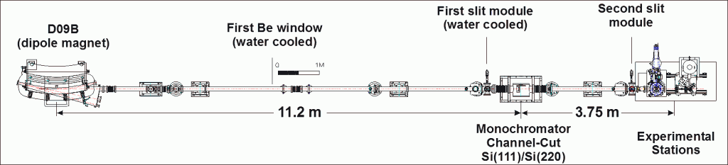
Português: Câmara de vácuo mostrando o arranjo de TXRF.
English:Vacuum chamber showing the TXRF setup.
Português:Porta-amostras do arranjo da microssonda de raios X.
English: Sample holder of the x-ray microprobe setup.
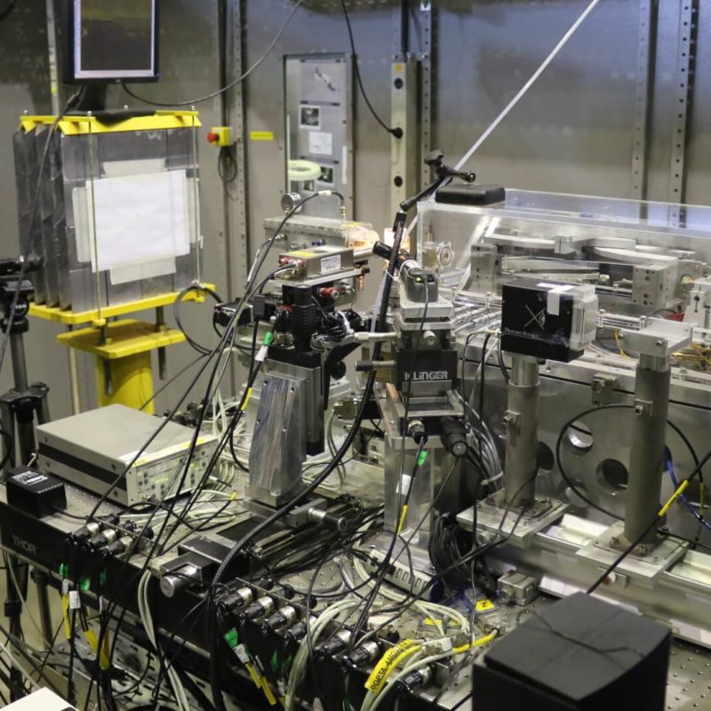
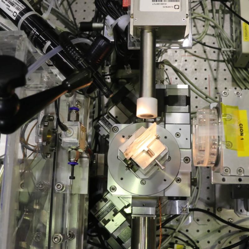
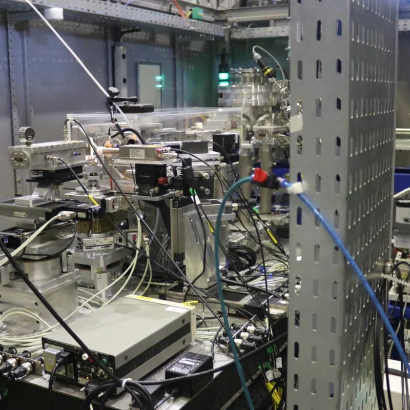
Português: Cabana experimental.
English: Experimental hutch
Português: Cabana experimental
English: Experimental hutch
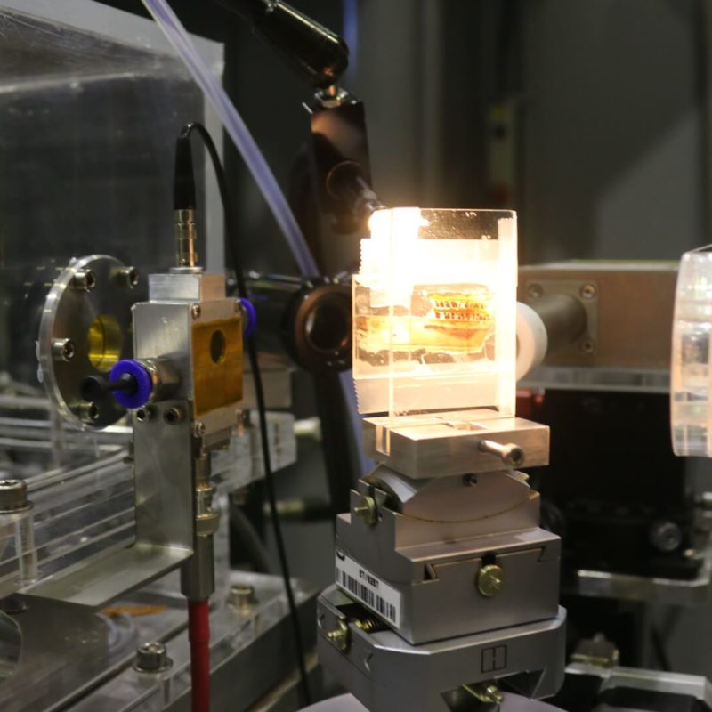
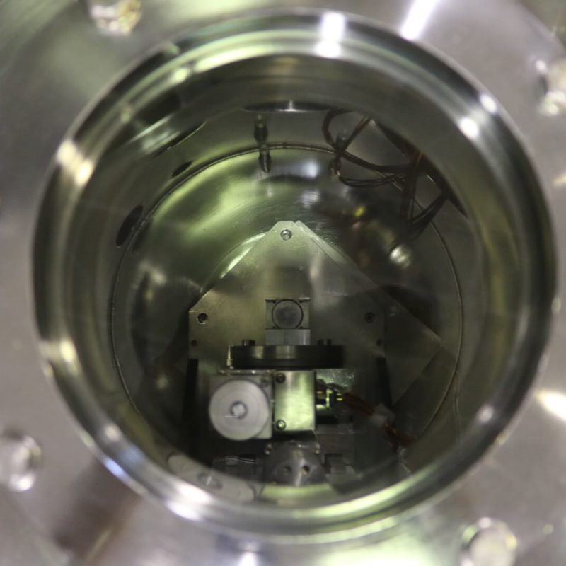
Português: Montagem de amostra no arranjo da microssonda de raios X.
English: Sample mounting on the x-ray microprobe setup.
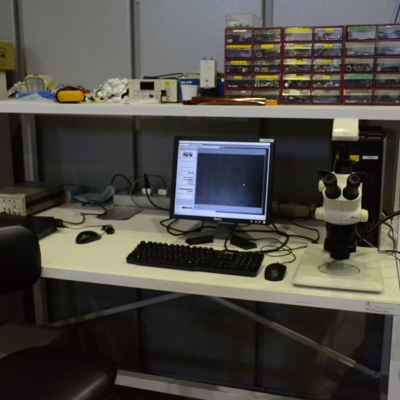
Português:Instalação de suporte para usuários.
English:User support facility.
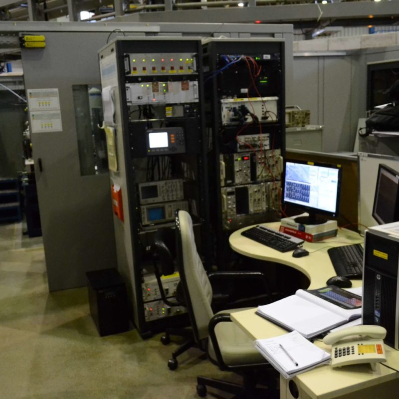
Português: Sala de controle da linha.
English: Beamline control room.