CONTACT & STAFF
Facility E-mail: jatoba@lnls.br


The JATOBÁ beamline is being built to produce a high-energy, high-photon flux beam focused on micrometric dimensions and will be dedicated to the study of a wide range of materials using the full X-ray scattering technique. The beamline should start users’ operation during the second semester of 2023.
The total scattering technique encompasses both Bragg diffraction from crystalline structures and diffuse scattering, which is related to short-range order effects and without the need for crystalline ordering. The signal obtained from the total scattering experiment is used to obtain the function known as PDF (Pair Distribution Function). The PDF function is an oscillatory function, and each peak represents in r (interatomic distance) the probability of finding a pair of atoms and is weighed by the scattering power of the pair of atoms [1]. Thus, based on total scattering experiments on the Jatobá beamline and the calculation of the PDF function, it will be possible to obtain information on the local order of amorphous and nanostructured materials, such as interatomic distances, degrees of disorder and coordination. [1].
JATOBÁ is essentially an X-ray scattering beamline, like PAINEIRA beamline for diffraction of polycrystals. However, to promote full X-ray scattering and PDF analysis experiments, it will deliver high energy X-rays, between 40 and 70 keV, corresponding to short wavelengths (0.3 to 0.17 Å), high Q (scattering vector modulus) of approximately 27 and 48 Å-1 and high photon flux (1012ph/s /100mA at 40keV) at the sample position. In addition, the beamline will be equiped with cell reaction, sample horder, gas-handlings systems and acessories similiar to the Paineira beamline. These infrastrutre will allow kinetcs experiments in in situ and operando conditions to study functional materials, such as catalysts, energy sotorage devices, for exemple.
[1] Egami, T. & Billinge, S. J. L. (2012). Underneath the Bragg Peaks: Structural Analysis of Complex Materials, 2nd ed. Amsterdam, Elsevier.
Facility E-mail: jatoba@lnls.br
X-ray diffraction (XRD) is one of the most established and used techniques among the several techniques that use synchrotron light. When reaching a crystal, X-ray photons are scattered by the electrons that make up the crystal and that are distributed periodically in the crystalline structure. This periodicity, of the same order as the X-ray wavelength, works as a diffraction grid that scatters the light in an inhomogeneous way. For some directions, the scattered light contributes in a constructive way generating what is called Bragg peak. The arrangement of these peaks, together with the incident beam energy, allows to determine the relative position of the atoms that make up the crystal. In the case of materials composed of a large number of theses crystals – usually of micro or nanometric dimensions composing a polycrystal – an average over all the polycrystalline material is made and the diffractogram appears, in the form of scattering rings around the direction of the incident beam. A finer modeling of these peaks allows to determine not only the crystalline phase, but also deformations in the crystalline network due to stress conditions and/or atomic inclusions.
Beyond collecting conventional diffraction patterns, as in the case of the PAINEIRA beamline, a unique capacity of JATOBÁ beamline is the possibility to probe a wider region of the reciprocal space, due to the high photon energy associated with large area detectors. In this technique, diffractograms resulting from X-ray interference patterns scattered in poorly ordered materials can be modeled to determine interatomic distances, giving information about the short and medium range order, complementary to long-range order of diffraction in polycrystals [1].
With an incident beam of micrometric dimensions, as is the case with JATOBÁ, it is possible to map regions with different crystallinities in the sample. Various types of analysis of nano-structured and / or poorly ordered materials will be possible through 2D and 3D mapping with micrometric spatial resolution and short, medium, and long-range order contrast. These images are obtained by means of high precision scanning in the positioning of the sample in relation to the micrometric beam of synchrotron light.
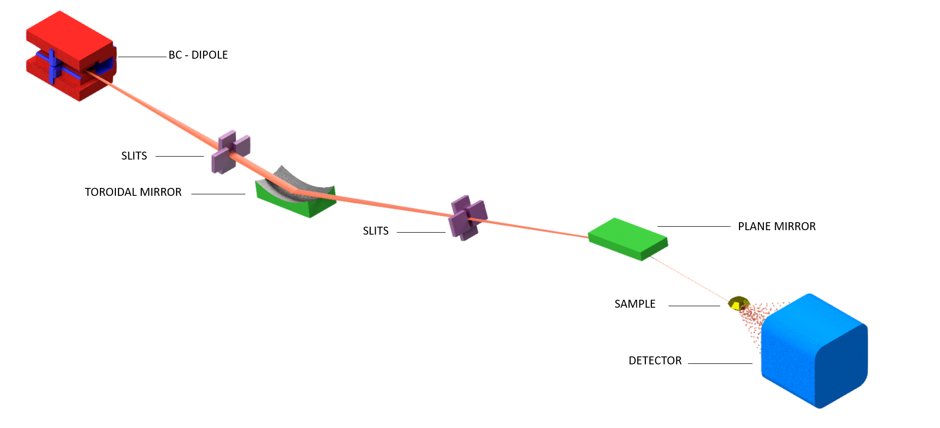
| Element | Type | Position [m] | Description |
|---|---|---|---|
| SOURCE | Dipole (BC) | 0 | BC |
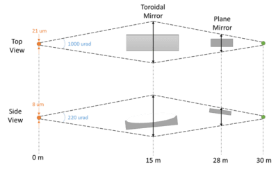
| ML1 | Multilayer Toroidal Mirror | 15 | Radiation extraction and focusing |
| ML2 | Multilayer Plane Mirror | 28 | Fine selection of energy |
| SE | Sample holder | 30 | Sample handler and environment |
| DET | Detector | 32 | 2D Detector |
The JATOBÁ beamline is designed to use the BC source by selecting three energies (E1, E2, E3) using multilayer mirrors.
| Parameter | E1 | E2 | E3 |
|---|---|---|---|
| Energy range [keV] | 43 | 57 | 71 |
| Wavelength [Å] | 0.29 | 0.22 | 0.17 |
| Qmax [Å-1] | 22 | 28 | 37 |
| Energy resolution (ΔE/E) [%] | 0.89 | 0.41 | 0.21 |
| Photon flux [photons/s/100mA] | 4.6 x 10¹² | 3.5 x 10¹² | 2.8 x 10¹¹ |
| Beam size [μm2, H x V – FWHM] | ~ 22 x 9.5 | ~ 22 x 9.5 | ~ 22 x 9.5 |