CONTACT & STAFF
Facility E-mail: sapucaia@lnls.br
Coordination: Aline R. Passos
Tel.: +55 19 3512 2332
E-mail: aline.passos@lnls.br
Click here for more information on this Facility team.


SAPUCAIA (Scattering APparatUs for Complex Applications and In-situ Assays) is a beamline dedicated to Small-Angle X-ray Scattering (SAXS), Ultra-Small-Angle X-ray Scattering (USAXS), and X-ray Photon Correlation Spectroscopy (XPCS). SAXS/USAXS are structural characterization techniques used to investigate properties such as shape, size, distribution, and spatial organization of objects at nano- and submicrometer scales. In addition to static measurements, these techniques enable structural changes to be monitored in situ and time-resolved kinetic studies to be performed with millisecond time resolution. XPCS complements this structural information by exploring temporal fluctuations in coherent X-ray scattering, allowing the investigation of dynamics, diffusion processes, and structural reorganization at nanometer and submicrometer scales. Together, these techniques enable the study of the structure and dynamics of complex systems, with applications in different areas of physics, chemistry, biology, materials science, and engineering.

Facility E-mail: sapucaia@lnls.br
Coordination: Aline R. Passos
Tel.: +55 19 3512 2332
E-mail: aline.passos@lnls.br
Click here for more information on this Facility team.
SAPUCAIA (Scattering APparatUs for Complex Applications and In-situ Assays) is a beamline dedicated to Small-Angle X-ray Scattering (SAXS) and Ultra-Small-Angle X-ray Scattering (USAXS). SAXS and USAXS are structural characterization techniques used to study properties such as shape, size, distribution, and spatial organization of nano- and microstructured objects. They also enable time-resolved kinetic studies with millisecond time resolution. These are well-established techniques with applications in diverse research fields, such as physics, chemistry, biology, and engineering. The beamline offers users the possibility of investigating relevant problems in the life sciences, with biological and medical applications, in structural biology, encompassing proteins, nucleic acids, lipids, and macromolecules in general, and in multiple topics in materials science, such as nanotechnology, polymers, and environmental sciences.
The SAPUCAIA beamline operates in a high-throughput regime, allowing rapid sample exchange. For this purpose, it has a robotic sample changer integrated with beamline control and data acquisition, enabling SAXS, USAXS, and XPCS experiments with high operational efficiency. The beamline is located in a high-beta section, ensuring low divergence. The source is a KYMA undulator, the beam is focused by a toroidal mirror, and the energy is defined by a DCM monochromator. The beamline is commissioned for the standard energy configuration of 8 keV. The PIMEGA 540D detector is installed in a vacuum chamber 15 meters long and 2 meters in diameter, allowing a sample-detector distance from 1 m to 10.5 m and a frame rate of 1 kHz, covering a wide q range. These properties enable the study of particles with dimensions ranging from a few nanometers to micrometers, making SAPUCAIA an extremely versatile tool for researchers from diverse scientific fields.
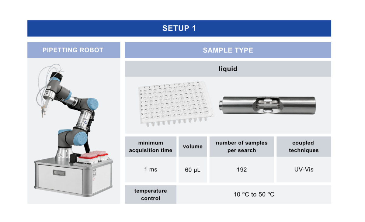
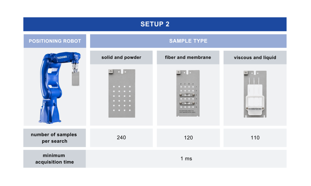
The SAPUCAIA beamline was designed to provide low parasitic scattering, low beam divergence, and high stability of the optical components. The experimental station has a robotic sample changer for liquids (with viscosity similar to that of water) and a dedicated positioner for solid samples (powder, film, fiber). The experimental configuration for liquid samples provides temperature control between 10 and 50 °C. In situ and time-resolved experiments can be performed, with time resolution down to 1 ms for TR-SAXS (Time-Resolved Small-Angle X-ray Scattering). For XPCS experiments, SAPUCAIA provides access to dynamic processes with characteristic times starting from the millisecond scale.
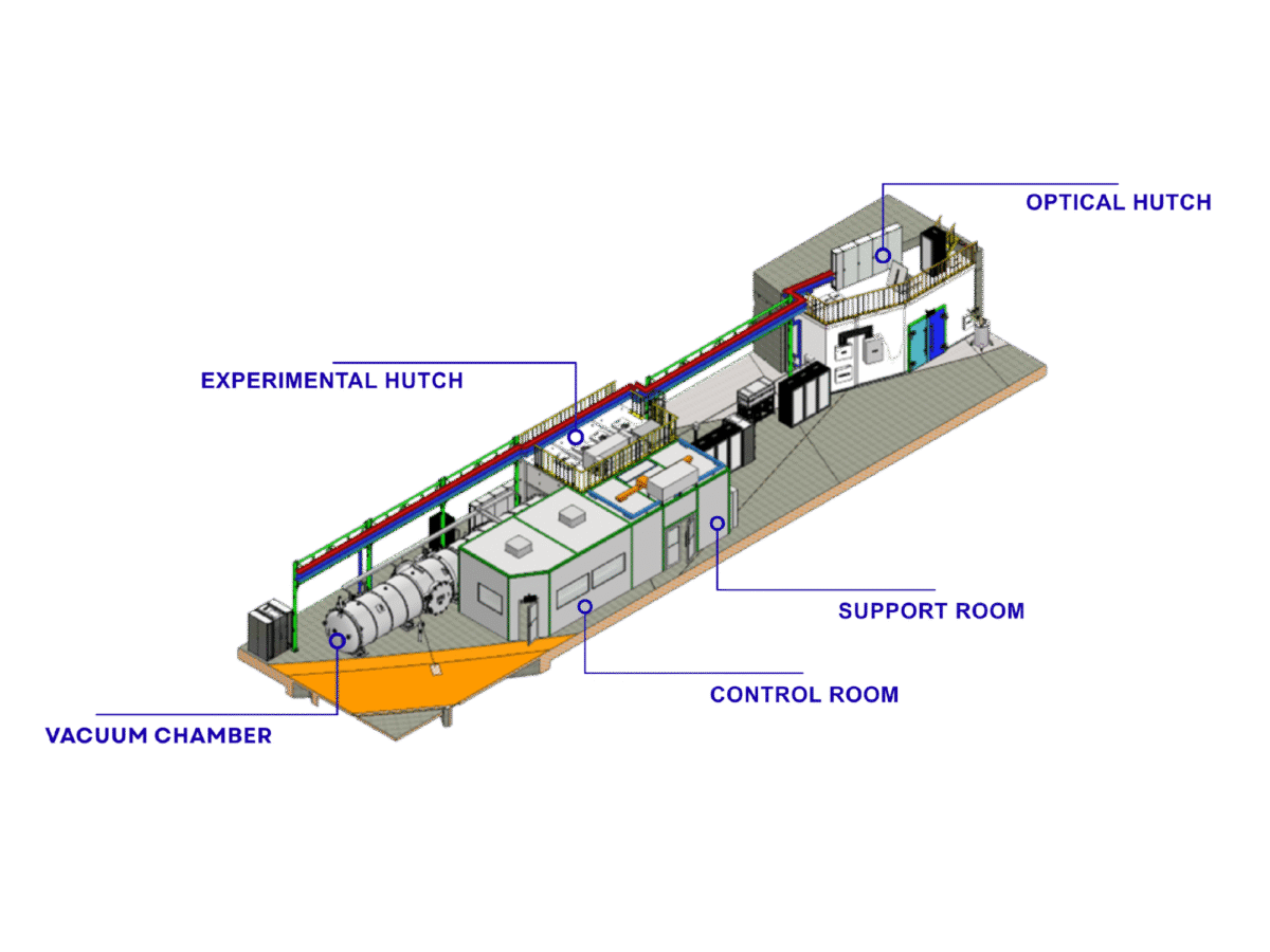
Representation of the SAPUCAIA beamline
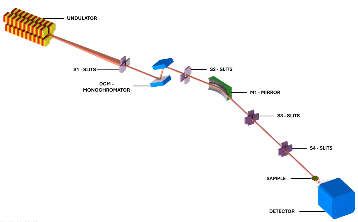
| Elements | Type | Position [m] | Description |
|---|---|---|---|
| SOURCE | Undulator | 0 | APU22 Kyma undulator, high beta straight section |
| S1 | White-beam slit | 27 | Slit located at the entrance of the optical hutch |
| DCM | Monochromator | 29 | Vertically deflecting double-crystal monochromator (Si 111 or Si 311) |
| S2 | Monochromatic-beam slit | 30 | Beam defining slits |
| M1 | Mirror | 31 | Toroidal Rh mirror (Meridional radius: 97 mm; Sagittal radius: 7900 m) |
| S3 | Defining slits | 32 | Beam-defining slits |
| S4 | Sample slits | 47.5 | Scatterless slits positioned in front of the sample |
| SH | Sample-holder | 47.8 | Sample-holder position |
| TN | Tunnel | 48 — 62 | Vacuum chamber with detector support |
| DET | Detector | 48.5 — 58.5 | PiMega 540D, Medipix3 |
| Parameter | Value |
|---|---|
| Energy range (keV) | 6 — 20 keV |
| Photon flux (photons/s/100mA) |
2 x 10¹² ph/s @ 8 keV |
| Photon flux (coherent mode) (photons/s/100mA) |
6 x 10¹⁰ ph/s @ 8 keV |
| Beam size at sample | ~ 200 µm2 |
| Sample-to-detector distance | 1 – 10.62 m |
| q-range | 0.004 – 5.2 nm-1 |
Small-Angle X-ray Scattering (SAXS) investigates the structural organization of materials at the nanometer scale through the elastic scattering of X-rays by variations in electron density present in the sample. These variations may occur, for example, between a particle and the solvent, between different domains of a material, or between distinct regions of a macromolecule. The scattered intensity therefore depends on the electron-density contrast between the structure of interest and the surrounding medium. Scattering is described in terms of the magnitude of the scattering vector, (q), which depends on the X-ray wavelength and the scattering angle. Since (q) is inversely related to the length scale being investigated, different regions of the SAXS curve correspond to different levels of structural organization: low (q) values identify larger structures, whereas higher (q) values reveal progressively smaller features.
Experimentally, the scattering pattern recorded on the detector is converted into an intensity curve I(q), which represents a statistical average of the structure of a large number of objects within the volume illuminated by the beam. The shape of this curve contains information about the dimensions, morphology, size distribution, internal structure, and spatial organization of the sample components. In particulate systems, its interpretation can be described in a simplified manner by the contributions of the form factor, P(q), related to the structure of the individual scattering objects, and the structure factor, S(q), associated with correlations and interactions between them. Data analysis, using model-independent approaches or structural models appropriate to the system, makes it possible to obtain parameters such as radius of gyration, characteristic dimensions, size distributions, distances between objects, and interface properties, as well as to monitor aggregation, self-assembly, and structural transitions.
For further study of SAXS/USAXS, consult the following references:
X-ray Photon Correlation Spectroscopy (XPCS) is a technique based on coherent X-ray scattering that enables the investigation of material dynamics at nano- and submicrometer scales. When a sample is illuminated with a coherent beam, the scattered waves interfere with one another and produce a granular intensity pattern on the detector, known as a speckle pattern. This pattern contains information about the instantaneous configuration of the sample and changes as its constituents move or reorganize. The temporal evolution of these fluctuations is quantified through the intensity autocorrelation function g₂(q, Δt), which compares the scattering pattern recorded at different times. The scattering vector q selects the spatial scale being investigated, while the interval Δt represents the temporal scale of the measurement. In this way, XPCS makes it possible to observe how structures at a given length scale remain correlated or reorganize over time.
The autocorrelation curves provide a characteristic relaxation time, whose dependence on q reveals information about the mechanism responsible for the system dynamics. For particles undergoing simple Brownian diffusion, for example, the relaxation rate Γ varies as q², following the relation Γ = Dq², where D is the translational diffusion coefficient. Other behaviors may indicate collective dynamics, confinement, heterogeneity, or out-of-equilibrium processes.
For further study of XPCS, consult the following references:
To obtain high-quality SAXS, USAXS, and XPCS data, the sample must provide an adequate scattering signal, remain stable during the measurement, and be compatible with the experimental environment used. In addition, the selected q range must be consistent with the size of the structures to be analyzed. Therefore, users should pay attention both to aspects related to the scattering from their samples, as well as to sample preparation and suitability for the experimental conditions offered by the SAPUCAIA beamline.
Regarding sample scattering, the user should consider:
Regarding the quality of sample preparation, the following factors should be kept in mind:
Regarding the suitability of the sample for the experimental conditions offered by the SAPUCAIA beamline:
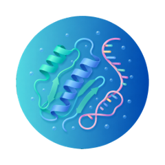
The function of biomacromolecules is closely linked to their organization in solution and their ability to transition between different structural states. Proteins, nucleic acids, and biomolecular complexes may exhibit multiple conformations, different oligomerization states, and transient associations that are not always accessible by methods based on isolated crystal structures. SAXS provides an integrated view of these structural populations in solution, enabling the investigation of conformation, flexibility, complex assembly, and equilibrium between different molecular states.
Conformational changes triggered by molecular interactions, pH, temperature, or solution composition can be systematically monitored, while time-resolved experiments extend these analyses to the evolution of association, dissociation, and structural reorganization processes. The high-throughput regime further extends this capability to condition screening and comparative studies involving large sets of samples.
XPCS extends this characterization by providing access to the collective dynamics of biomolecular systems, particularly in concentrated solutions and systems exhibiting supramolecular organization. The technique can monitor processes such as diffusion, phase separation, condensate formation, gelation, and viscoelastic relaxations, enabling investigation of how the mobility and interactions of biomacromolecules evolve over time and across different spatial scales.
Related publications:
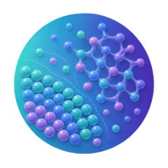
In colloidal systems, the relevant structure lies not only in the morphology of each particle, but also in the way their constituents interact and organize collectively. Small variations in interparticle interactions can produce profound changes in system behavior, leading to the formation of aggregates, percolated networks, gels, glasses, colloidal crystals, and other organized phases. SAXS and USAXS provide direct access to this organization over a broad range of scales, connecting the structure of individual units to the hierarchical architectures that emerge in concentrated and complex systems.
This approach is particularly powerful for investigating how structure forms and evolves, rather than only its final state. In situ and time-resolved measurements can follow self-assembly pathways, nucleation and phase growth, structural transitions, gelation, and aging, revealing intermediate states and out-of-equilibrium structures. The possibility of exploring nanometer to submicrometer scales makes SAXS/USAXS especially suitable for relating microscopic interactions to mesoscopic organization and, ultimately, to the macroscopic properties of colloids and soft-matter materials.
Dynamics is a fundamental component of the behavior of colloidal systems and can be investigated directly by XPCS. The technique makes it possible to quantify diffusive and collective motions, relaxation times, aging, and responses to external perturbations, as well as to follow the evolution of out-of-equilibrium systems. The combination of SAXS/USAXS and XPCS therefore makes it possible to correlate the structural organization of colloids with the dynamic processes responsible for their evolution.
Related publications:

Nanomaterials developed for medical applications are systems whose structure can be profoundly modified by the environment in which they are found. The encapsulation of therapeutic molecules, changes in medium composition, and interactions with proteins, lipids, and membranes can alter their internal organization, stability, and association state. Understanding these transformations is essential for establishing relationships between nanomaterial architecture and biological performance.
SAXS makes it possible to investigate nanocarriers directly in solution and to monitor structural changes associated with formulation, encapsulation, and release of molecules. At nano-biological interfaces, the technique provides a particularly valuable perspective by enabling observation of changes that arise after nanoparticles come into contact with the biological environment, including biomolecular corona formation, structural reorganization, and aggregation. This capability makes it possible to compare the originally designed structure with that actually exhibited by the nanomaterial under biologically relevant conditions, providing fundamental information for the rational development of lipid nanoparticles, liposomes, polymeric systems, and inorganic nanomaterials intended for biomedical applications.
XPCS makes it possible to investigate in situ the dynamics of nanoparticles in complex biological environments, following changes in their diffusion arising from interactions with biomolecules and with the surrounding medium. Changes in nanoparticle mobility can reveal processes such as protein corona formation, aggregation, and changes in stability, enabling evaluation of the evolution of the nano-biological interface directly in concentrated and highly complex media.
Related publications:
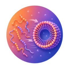
In polymeric materials, small changes in molecular architecture or processing conditions can produce profound changes in nanoscale organization. Block copolymers, polymeric particles, gels, and hybrid materials can form domains, networks, and periodic structures whose organization determines mechanical, optical, transport, and stimulus-response properties. SAXS and USAXS make it possible to follow this organization across different scales and establish relationships between composition, processing, and final structure.
One of the most powerful applications of SAXS in this area is observing material formation as it occurs. Time-resolved SAXS can directly follow particle nucleation and growth, polymerization, phase separation, and polymerization-induced self-assembly processes, revealing structural intermediates that are no longer present in the final product. In self-assembled materials, the evolution of scattering patterns makes it possible to identify the emergence and transformation of ordered phases, providing a direct view of how structure emerges and reorganizes during synthesis, processing, or the application of external stimuli.
In polymeric and self-assembled materials, XPCS provides access to the microscopic relaxations that accompany the formation and reorganization of ordered structures. The technique can reveal collective motions, domain rearrangements, and differences in dynamics between different phases, complementing the structural information obtained by SAXS and enabling the architecture of the material to be related to the mechanisms responsible for its temporal evolution.
Related publications:
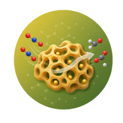
Catalysts and porous materials are often designed from structures that extend across different length scales. Active nanoparticles, aggregates, interfaces, pores, and hierarchical structures may evolve during synthesis, activation, and operation, directly altering the activity, selectivity, and stability of the material. SAXS and USAXS provide a way to follow these transformations at nanometer and mesoscopic scales, including in heterogeneous materials in which the relevant structure is not restricted to the crystalline phase.
This approach becomes especially important in in situ and operando experiments, in which the structure can be observed during material operation. Nanoparticle growth and coalescence, sintering, morphological reconstructions, and degradation processes can be followed over time and correlated with reaction conditions. In porous materials and frameworks, time-resolved measurements can also reveal intermediates and organizational steps that precede formation of the final structure. Combined with techniques sensitive to crystalline structure or chemical state, SAXS thus provides a multiscale description of the evolution of catalysts and functional materials under relevant operating conditions.
XPCS adds a temporal dimension to the study of porous materials by enabling fluctuations, reorganizations, and growth processes to be followed under in situ conditions. In systems such as zeolites, analysis of temporal scattering correlations can distinguish different stages of nucleation, growth, phase transformation, and crystallization, providing dynamic information that complements the structural evolution observed by SAXS.
Related publications: