CONTACT & STAFF
Facility E-mail: cedro@lnls.br
Coordination: Juliana S. Yoneda
Tel.: +55 19 3512 1045
E-mail: juliana.yoneda@lnls.br
Click here for more information on this Facility team.


CeDRO (Circular Dichroism Beamline) is one beamline of the Brazilian Synchrotron Light Laboratory (LNLS) in the commissioning phase at the Sirius storage ring. CEDRO is dedicated to the electronic circular dichroism (CD) spectroscopy in the ultraviolet region (UV). CD spectroscopy is based on the differential absorption of circularly polarized light to left and right by optically active entities, i.e., chiral materials. It is a valuable tool to analyze the structure of chiral molecules, including biomolecules such as proteins, nucleic acids, and carbohydrates. Amongst them, the proteins are extensively studied by CD to quantify the secondary structure content in terms of α-helical, β-strand, unordered and other structure and to study folding, stability, and interactions in solution, which are significant in the search and development of new drugs, for instance.
Facility E-mail: cedro@lnls.br
Coordination: Juliana S. Yoneda
Tel.: +55 19 3512 1045
E-mail: juliana.yoneda@lnls.br
Click here for more information on this Facility team.
⚠ The CEDRO beamline is operating in Fast Track mode.
To perform circular dichroism measurements in the CEDRO beamline, we have a CD spectrometer that is being coupled to the UV synchrotron beam. Currently (2nd semester 2023), we are not yet in the ideal condition, therefore, the beamline is not 100% operational and is operating in hybrid mode: measurements can be carried out using the conventional source (UV lamp) and the synchrotron beam, although this last is not fully optimized.
Usual measurements, such as for samples in solution, can be carried out, including temperature control/ramping, using different pathlengths quartz cells. Quartz plates are also available for thin film samples. Special sample holders (with automatic rotation and translation to change the sample’s position in relation to the detector) are being developed and will soon be made available to users who need these requirements.
Please, contact the beamline team if you have any questions before submitting your proposal: cedro@lnls.br


M4 will be used in the future to change the direction of the beam to generate a second branch for an additional experiment.

CEDRO beamline is comprised by an experimental station to perform SRCD experiments. Sample holders for liquid samples with temperature control are available. Another branch with a second experiment is foreseen in the future. There is a control room, where the users can control, follow the measurements, and perform pre analysis while experiments are running. An ancillary laboratory is also available for sample handling with some simple equipment, such as thermal bath, vortex, mini centrifuge, refrigerated centrifuge, Nanodrop, pHmeter, balance, heating plate, fridge and freezer.

Overview of the CEDRO experimental station, control room and ancillary laboratory.
The technique is called SRCD (Synchrotron Radiation Circular Dichroism) when the light source is the synchrotron radiation (SR). SR offers enhanced photon flux compared to conventional lamps used in laboratory-based instruments increasing the sensitivity and, it enables data collection at lower wavelengths, in the vacuum ultraviolet region (VUV, below 200 nm), which is commonly inaccessible in a conventional instrument [1].
The extension of wavelength range to the VUV region allows the detection of additional high-energy transitions. For instance, the n–σ* transitions of acetal and hydroxyl groups of saccharides produce bands at 140–180 nm [2]. Further, the protein secondary structure content can be quantified, and this analysis is improved with the increase of spectrum wavelength range. The protein CD spectra can be considered as the weighted sum of the secondary structure components. The amount of information that can be extracted depends on the number of transitions included in the spectrum. It is known that spectra achieving wavelengths not lower than 200 nm can be described by two eigenvectors allowing the estimation of helices, only. This value increases to three or four when the spectrum is extended to 190 nm and six to eight to 170 nm, enabling the distinction among a greater number of secondary structures [3]. While the increased intensity of incident light is crucial for enhancing the signal-to-noise ratio, it is essential to set limits, as excessive exposure may lead to radiation damage [4]. By the other hand, the high photon flux can be employed in experiments involving UV-induced denaturation, as demonstrated in the SRCD beamline (B23) at Diamond [5]. In summary, the use of synchrotron radiation as the light source enhances the utility of CD spectroscopy.
[1] BA Wallace. The role of circular dichroism spectroscopy in the era of integrative structural biology. Current Opinion in Structural Biology. 2019; 58:191–196.
[2] K Gekko. Synchrotron-radiation vacuum-ultraviolet circular dichroism spectroscopy in structural biology: an overview. Biophys Physicobiol. 2019; 16: 41–58.
[3] Lees JG, Miles AJ, Wien F, Wallace BA. A reference database for circular dichroism spectroscopy covering fold and secondary structure space. Bioinformatics. 2006; 22:1955e62.
[4] AJ Miles, RW Janes, A Brown, DT Clarke, JC Sutherland, Y Tao, BA Wallace, SV Hoffmann. Light flux density threshold at which protein denaturation is induced by synchrotron radiation circular dichroism beamlines. J. Synchrotron Rad. 2008),15, 420–422.
[5] R Hussain, E Longo, G Siligardi. UV-Denaturation Assay to Assess Protein Photostability and Ligand-Binding Interactions Using the High Photon Flux of Diamond B23 Beamline for SRCD. Molecules 2018, 23, 1906.

The incoming beam from a bending magnet (B2 dipole) goes through optics elements before reaching the monochromator (Hummingbird subtractive double grating – OLIS®) (drawing is for illustrative purposes only). A CaF2 window between the optics chamber and the monochromator separates the UHV (ultra-high vacuum) region from the N2 purged atmosphere. The monochromatized light pass through a Rochon Polarizer to ensure the linear polarization of the beam. The linear polarized light (LPL) is converted to circularly polarized light (CPL) alternatively to right and left by a photoelastic modulator and pass through the sample before reaching the detector (photomultiplier tube). The signal is digitalized, and the CD spectra are obtained after digital processing.
Sample preparation is an important step, and the choice of sample conditions is a key factor to obtain good CD results. As CD spectroscopy relies on the differential absorption of a chiral sample, care must be taken about the overall absorption of the sample. Components that absorb strongly in the wavelength region of interest must be avoided or kept at very low concentration for obtaining a spectrum with a good signal-to-noise level.
Some tips about solvent and buffers:
Phosphate, Tris, and Borate are suitable for CD studies (260 – 180nm). Chloride ions need to be avoided, as it absorbs strongly below 200 nm, therefore be aware when using HCl to adjust the required pH and, as the component in the phosphate buffered saline (PBS). If the presence of salts is needed to maintain the ionic strength, the use of sulphate or fluoride is preferably. HEPES, MOPS, MES, PIPES should be used only at low concentrations. Imidazole, which is used to elute His-tagged proteins from immobilized metal chromatography, should be removed by extensive dialysis or gel permeation.
If the biological requirements make difficult the replacement of certain components, using a cell of very short pathlength can mitigate the problem caused by the high absorbance of the buffer. In this case, the protein concentration should be higher, therefore, be aware whether the increase of the protein concentration is not causing aggregation or precipitation.
One good advice is running some initial tests to find the best sample cell pathlength and instrument parameters.
We recommend reading articles on experimental conditions to better plan your CD studies:
For liquid samples we have two options of cells. The pathlength can be chosen according to protein concentration. The Table below can aid your choice.

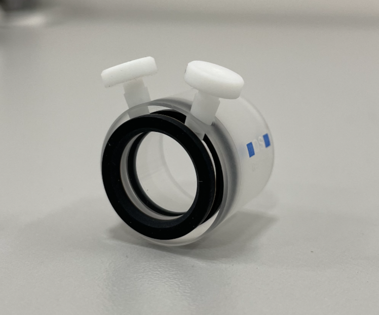
Closed Suprasil Quartz round cells with Teflon stoppers (Hellma 121.000)
This type of cell is recommended for studies with temperature change of the sample (thermal stability experiments). The pathlengths available are 0.1, 0.2 and 0.5 mm.
The cleaning can be problematic for these cells with very short pathlengths, and care must be taken in this process. Please, ask for assistance if you need.
Demountable Suprasil quartz round cells (Hellma 124.000)
Two demountable plates, one is flat and the another has a beveled edge producing a thin well. Typical pathlengths are between 0.01 and 0.1 mm.
The cleaning is easier because they are separated in two plates, however the loading can be more laborious. Practicing (e.g., with water) before loading the sample is convenient for those who have not yet used this type of cell. Please, ask for assistance if you need.

Suprasil quartz plates (Hellma 202)
Suprasil quartz plates can be used as substrates for film preparation. The construction of a sample holder with humidity control and rotation stage is planned for measurements in solid phase (dehydrated films) for oriented CD experiments, for instance. It should be stressed when measuring solid sample is necessary to control the interference of linear dichroism due to possible sample alignment upon drying. One can perform the measurements at different rotation angles around the beam propagation axis.

Microfluidic devices will be developed envisaging kinetic studies.

The electronic transitions in the backbone peptide bond of proteins occur in the far UV region (160 to 240 nm). The n→π* and π*→π* transitions of the amide chromophore are influenced by the geometry of the polypeptide backbones; therefore, CD spectra are sensitive to protein secondary structures. The spectra can be deconvoluted to quantify the contributions of different types of secondary structure in a protein. Different methods have been developed to perform this analysis. One can use a reference set composed by spectra of proteins with known 3D structure (e.g., by crystallography and X-ray diffractions or RMN) and different empirical algorithms.
B.A. Wallace. Protein characterisation by synchrotron radiation circular dichroism spectroscopy. (2009) Quarterly Reviews of Biophysics , Volume 42 , Issue 4 , pp. 317 – 370. DOI: 10.1017/S003358351000003X

The dehydration of a protein solution forming a thin film allows the CD measurements below ~168 nm, which is normally the lowest cutoff to solutions due to the water absorption of light at that wavelength. The extension of energy range enables the detection of additional transitions increasing the amount of information that can be extracted from the CD spectrum.
Yoneda JS, Miles AJ, Araujo AP, Wallace BA. (2017) Protein Science, 26(4):718-726. doi: 10.1002/pro.3118

Thermal denaturation can be induced to evaluate the protein stability. For instance, the melting temperature of a protein can be compared at different conditions to detect changes in its stability. Further, the thermal denaturation can be used to detect ligand binding, as this interaction can cause changes in protein stability.
Lopes JLS, Yoneda JS, Martins JM, DeMarco R, Jameson DM, Castro AM, et al. (2016) Environmental Factors Modulating the Stability and Enzymatic Activity of the Petrotoga mobilis Esterase (PmEst). PLoS ONE 11(6): e0158146. DOI: 10.1371/journal.pone.0158146

Oriented circular dichroism (OCD) can be used to address the alignment of α-helical peptides related to lipid membranes. Moffitt’s theory predicts that the electronic transition dipole moments of the backbone amide bonds in helical polypeptides are polarized either parallel or perpendicular to the helix axis. The exciton splitting of the π – π* transition centered around 200 nm results in vector components parallel and perpendicular to the helix axis, giving rise to a negative 208 nm peak and a positive 190 nm peak. On contrary to isotropic solutions where proteins can assume all the orientations related to the light beam, in an anisotropic sample, the OCD spectrum of an α-helical peptide interacting with lipid membrane exhibits a characteristic line shape which reflects the angle between the helix axis and the lipid bilayer normal.
*Figure inspired from Bürck et al, 2016.
Bürck J, Wadhwani P, Fanghänel S, Ulrich AS. Oriented Circular Dichroism: A Method to Characterize Membrane-
Active Peptides in Oriented Lipid Bilayers (2016) Acc Chem Res. 49(2):184-92. DOI: 10.1021/acs.accounts.5b00346A. J. Miles, and B. A. Wallace. Circular dichroism spectroscopy of membrane proteins (2016) Chem. Soc. Rev., 2016,45, 4859-4872. DOI: 10.1039/C5CS00084J