CONTACT & STAFF
Facility Tel.: +55 19 3518 2364
Facility E-mail: carnauba@lnls.br
Coordination: Rodrigo Szostak
Tel.: +55 19 3517 5156
E-mail: rodrigo.szostak@lnls.br
Click here for more information on this Facility team.


CARNAÚBA (Coherent X-rAy NAnoprobe BeAmline) is the longest beamline of the Sirius with approximately 145 meters between the light source and the sample environment, which allows a high optical demagnification and to reach nanometric spatial resolutions. It has two endstation: TARUMÃ (Tender-to-hard X-ray for sub-micro analysis), with submicrometric beam size and variable sample environment; and the SAPOTI (Scanning Analysis by Ptycho for Tomographic Imaging) in which the beam size reaches 30 nm and the sample environment is cryogenic and in utra-high vacuum.
The CARNAÚBA beamline covers the energy range from 2.05 to 15 keV and it comprises multiple techniques based on X-ray absorption, scattering and emission. In this beamline, it is possible to access the K edges of light elements, such as phosphorus and sulfur, which are quite relevant for Life and Environmental Sciences and also L-edges of elements of technological importance, such as the Lanthanoids. It is possible to study many types of nano-structured materials and hierarchically ordered ones through 2D and 3D images using as contrast X-ray absorption, emission, and diffraction, as well as optical emission. Some of the main areas which benefit from these techniques are: Materials Science (catalysts, magnetism, semiconductors, electrochemistry, photonics); Nanotechnology (health, information); Environmental Sciences (geosciences, materials under extreme pressure, petrology); Cultural Heritage (arts, archeology and paleontology) and Life sciences (medical and biological applications).
The optical design of this beamline takes advantage of the Sirius low emittance, allowing the beam at the sample to be nanometric and relatively low divergence at the same time with a large depth of focus. The later is an important aspect for the phase contrast diffraction technique.
⚠ FFor cycle 7 (Scheduling period august 2026 to February 2027), the CARNAUBA beamline will operate from 2-15 keV for excitation energies. The experiments CNBT_EXP13 and CNBT_EXP14 run over the energy range 3-6 keV under standard conditions with EVL=3. However, experiments with excitation energies between 2 and 3 keV are also possible, provided they are well discussed with the beamline staff. We advise that experiments in the range of excitation energy of 2-3 keV are more challenging and risky. We advise users to contact the beamline staff (by e-mail) to discuss the feasibility and to pre-submit the proposals as soon as possible.
Facility Tel.: +55 19 3518 2364
Facility E-mail: carnauba@lnls.br
Coordination: Rodrigo Szostak
Tel.: +55 19 3517 5156
E-mail: rodrigo.szostak@lnls.br
Click here for more information on this Facility team.
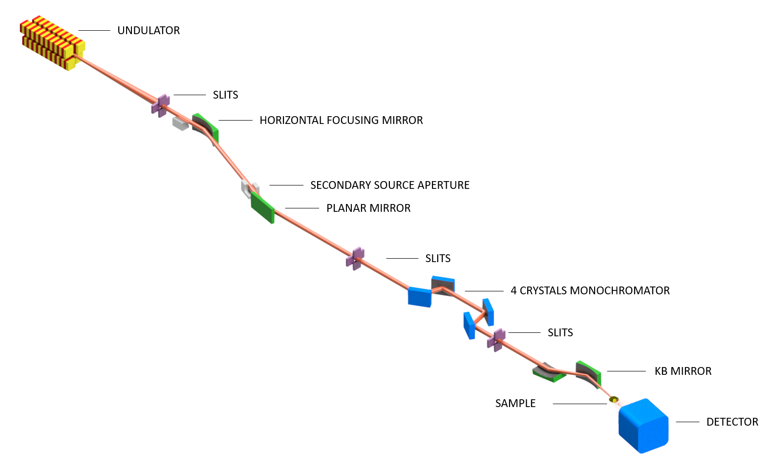
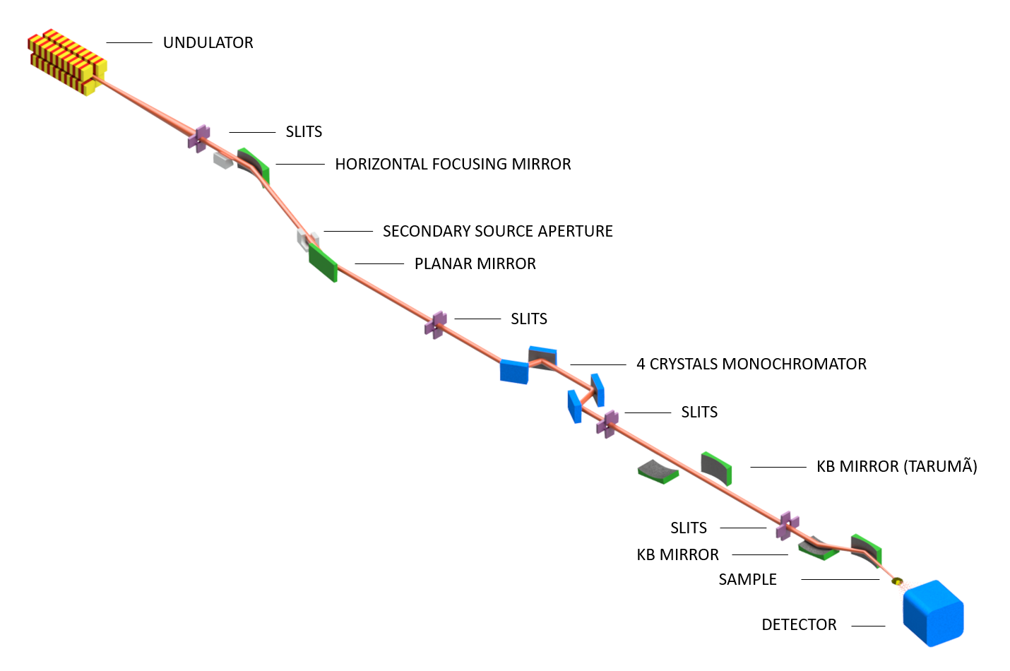
| Element | Type | Position [m] | Description |
|---|---|---|---|
| SOURCE | Insertion Device | 0.0 | Delta Undulator |
| M1 | First Mirror | 27.4 | Focusing |
| SSA | Secondary Source | 54.0 | Defining source aperture |
| M2 | Second Mirror | 54.3 | Horizontal deflection |
| 4CM | Four-bounce Monochromator | 130.0 | Monochromatization |
| KB Mv Tarumã | Vertical KB Mirror | 134.2 | Vertical Focusing |
| KB Mh Tarumã | Horizontal KB Mirror | 134.5 | Horizontal Focusing |
| Sample Tarumã | Sample position | 135.0 | |
| KB Mv Sapoti | Vertical KB Mirror | 142.6 | Vertical Focusing |
| KB Mh Sapoti | Horizontal KB Mirror | 142.9 | Horizontal Focusing |
| Sample Sapoti | Sample position | 143.0 |
The TARUMA (Tender to hArd x-ray foR sUbMicro Analysis) nanoprobe station started to operate in November 2020 for technical and internal scientific commissioning and is currently fully operational for standard calls for proposals. More details about the CARNAÚBA beamline and TARUMÃ station can be found in the reference https://www.sciencedirect.com/science/article/pii/S0368204823000579.
The SAPOTI end station is under assembly. It is expected to start technical commissioning in 2025. More details about the SAPOTI can be found at https://pubs.aip.org/aip/acp/article/2990/1/040017/2913068/The-high-dynamic-cryogenic-sample-stage-for-SAPOTI.
| Parameter | Value | Condition |
|---|---|---|
| Energy Range | 2.05 – 15 keV | Si(111) |
| Energy Resolution (ΔE/E) | 10-4 – 10-5 | |
| Harmonic Content | < 10-5 | Above 5 keV |
| Energy Scan | Yes | |
| Beamsize at sample [μm] @Tarumã | 0.15 x 0.15 (0.55 x 0.55) | 8 keV (2 keV) |
| Beam Divergence at sample [mrad] @Tarumã | (1 x 1) | All energy range |
| Estimated flux [ph/s/100 mA] @Tarumã | 1011 | – |
| Beamsize at sample [μm] @Sapoti | 0.03 x 0.03 (0.12 x 0.12) | 8 keV (2 keV) |
| Beam Divergence at sample [mrad] @Sapoti | 5×5 (4 x 4) | < 10 keV (12keV) |
| Estimated flux [ph/s/100 mA] @Sapoti | 1012 | – |
| Imaging Mode | Scanning | – |
| Coherence Modes | ~1 | – |
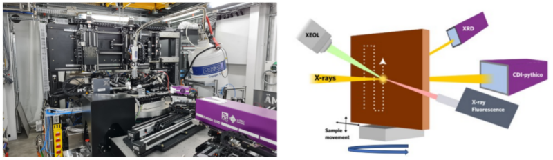
The TARUMA end station is equipped with translation and rotation stages that allow the raster scan while collecting the signal in different detectors. The XYZ piezo stage allows nanometer-level 2D mapping via fly-scan mode. The Figure below shows a simple schematic view of the beamline with the sample and detector arrangement. Depending on the measurement conditions, more than one technique can be used simultaneously.
X-rays are electromagnetic radiation that, upon interaction with matter, produce secondary radiation carrying useful information about the atoms that make up the target. Photons can interact with matter in different ways depending on their energy. Photons in the X-ray regime interact with the electron shells that surround the nucleus. There are three photon-atom processes whose influence prevails in the X-ray regime: The Compton scattering, the Rayleigh scattering, and the photoelectric effect. For the latter, photons cause the ejection of a core electron, leaving a hole in the atom. Then, the electron hole is filled by an electron from an outer shell, emitting a fluorescence photon with an energy characteristic of each atom and transition. This fluorescence emission allows a qualitative and quantitative determination of the elemental composition of the material. By raster scanning the sample using the nanobeam available at CARNAÚBA beamline, one can obtain hyperspectral images (2D XRF maps) containing chemical information of the sample with nanometric spatial resolution.
Crystalline material is composed of atoms ordered periodically that scatter X-rays in very specific directions to produce a scattering pattern with sharp maxima, the diffraction peaks. In an X-ray diffraction (XRD) pattern, the diffraction peaks have positions related to the distance between the atoms (scatters) mathematically represented by the well-known Bragg Law. An XRD pattern can provide information about the structure, phases, preferred crystal orientations (texture), and other structural parameters, such as average grain size, crystallinity, strain, and crystal defects. In synchrotron facilities with a nanofocus beam, the nanodiffraction (Nano-XRD) can be obtained. The XRD pattern is obtained from a small spot with nano to micrometric resolution. It allows for the taking of information about a determined local area that is different from an average area when a conventional beam (millimeter) is used. When combined with a scanning stage, a 2D XRD map can be obtained and give spatial information about the local crystal structure. The Carnauba beamline is equipped with an area detector, allowing nano-XRD experiments.
The XAS technique is based on the irradiation of the sample by a monochromatic X-ray beam. Then, the absorption of these photons is measured as a function of the incident photon energy. In XAS, an element-specific absorption edge is selected, allowing the study of isolated elements inside a sample. XAS spectra can be divided into X-ray absorption near-edge spectroscopy (XANES) and Extended X-ray absorption fine structure (EXAFS). XANES is strongly sensitive to the formal oxidation state and the coordination chemistry of the absorbing atom, while EXAFS is used to determine the distances, coordination numbers, and species of the neighbors of the absorbing atom. In Carnauba, the XANES can be measured in fluorescence or transmission mode in punctual or by obtaining XANES maps, both with spatial resolution on the range of the beam size.
X-ray ptychography is a lensless imaging technique that uses a coherent X-ray beam to scan overlapping regions of a sample, collecting diffraction patterns that are computationally reconstructed into high-resolution images. By retrieving both the amplitude and phase of the scattered waves, it provides detailed structural, compositional, and phase contrast information, making it ideal for imaging weakly absorbing samples like biological tissues or nanostructures. This method can achieve resolutions at the nanometer scale and, when combined with tomography, enables 3D visualization of internal structures. Its non-destructive nature, coupled with the ability to reveal electronic and elemental properties, makes ptychography a powerful tool in materials science, biology, and semiconductor research. In Carnauba, the ptychography can be measured simultaneously to fluorescence and transmission maps, which enables precise correlations between nanoscale structural features and elemental composition, providing a comprehensive view of materials’ morphology, chemical distribution, and heterogeneity.
Bragg Coherent Diffraction Imaging (BCDI) is an advanced imaging technique that utilizes coherent X-rays to investigate the morphology and internal properties of single nanoparticles. This method is based on collecting coherent diffraction patterns around a Bragg peak, which are subsequently processed using computational algorithms to generate three-dimensional images of the electron density and strain field of the crystal. BCDI is particularly powerful for studying defects, internal stresses, and structural dynamics in materials with nanometric resolution. Its applications include the investigation of functional materials such as catalysts and batteries, where analyzing structural changes under operational conditions (in situ or operando) is essential, enabling a detailed understanding of processes that influence the performance and durability of these materials.
Luminescence is a general term used to describe the spontaneous emission of light following an external stimulus. In X-ray excited optical luminescence (XEOL), this emission occurs after the absorption of X-rays, followed by the rearrangement of charge carriers in the material’s electronic structure. This process results in the recombination of electron-hole pairs and the subsequent emission of light, which can range from ultraviolet to near-infrared.
XEOL is a photon-in / photon-out technique that can be analyzed in both the time and energy domains. At Carnaúba, optical contrast has been explored in studies involving energy scanning, multispectral acquisitions, and 2D hyperspectral imaging. Additionally, XEOL can be combined with other techniques available on the beamline, enabling the correlation of the optical properties of different materials with the evaluation of their crystalline structure, chemical composition, and morphology.
The STXM technique enables the acquisition of two-dimensional images with high spatial resolution, using X-ray absorption as the contrast mechanism. In this method, the sample moves in front of a nanometric X-ray beam, while the transmitted X-ray flux is recorded by a photodiode at each position reached by the beam. By tuning the energy to the absorption edge of a specific element, it is possible to perform spectromicroscopy. This allows images obtained in very small regions to display contrast based on both elemental composition and oxidation state.
When fluorescence and absorption tomographies are performed simultaneously using scanning nanoprobes like those at the CARNAÚBA beamline, a unique and powerful opportunity arises to study materials with both structural and chemical detail in 3D. Fluorescence tomography provides precise elemental mapping, identifying the spatial distribution of specific elements within a material, while absorption tomography offers insight into the material’s density and internal structure. By combining both techniques, researchers can correlate elemental composition with structural features, enhancing the understanding of complex materials and systems. Furthermore, ptychography-tomography can be integrated to achieve extremely high resolution, revealing nanoscale details such as defects, interfaces, and fine structural variations. As a result, this combined approach allows for a comprehensive 3D visualization of both the chemical and morphological features of the material.
The CARNAÚBA is equipped with several sample environments and holders to serve the community. Below are the ones currently available. Do not hesitate to contact the beamline staff to get more details before submitting the proposal.
![]()
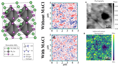
Climate changes have motivated scientists to find materials to improve energy conversion technologies. In this context, solar cells, especially perovskite solar cells (PSCs), have caught the attention and are promising for massive commercialization soon. Metal halide perovskites (MHP), the active layer of the PSCs, are composed of a mixture of Cs, MA, and FA at site A, I, or Br at site X, and Pb at site B. This material presents appropriate properties to solar cells, that result in power conversion efficiency similar to silicon solar cells in lab scale. Despite being synthesized with controlled methods, these materials present chemical, structural, and optical heterogeneities on the nanoscale. The Carnauba beamline has been used to investigate halide heterogeneities in MHP through the nano-XRF, revealing the effect of the additives to improve homogeneity. Besides that, the damage caused by the X-ray, as well as the strategies to mitigate it and find appropriate conditions to use X-ray ptycography, also has been investigated. The beamline is equipped with a photovoltaic holder that can be used for in situ and operando studies.
da Silva, F. M. C.; Szostak, R.; Guaita, M. G. D.; Teixeira, V. C.; Nogueira, A. F.; Tolentino, H. C. N. X-ray dose effects and strategies to mitigate beam damage in metal halide perovskites under high brilliance X-ray photon sources. Energy Mater. 2024, 4, 400058. http://dx.doi.org/10.20517/energymater.2023.114
G. D. Guaita, R. Szostak, F. M. C. da Silva, A. de Morais, R. F. Moral, T. Kodalle, V. C. Teixeira, C. M. Sutter-Fella, H. C. N. Tolentino, A. F. Nogueira, Influence of Methylammonium Chloride on Wide-Bandgap Halide Perovskites Films for Solar Cells. Adv. Funct. Mater. 2024, 34, 2307104. https://doi.org/10.1002/adfm.202307104
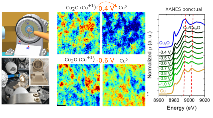
Electrochemistry is undeniably important in modern society as it directly impacts major technological and economic sectors such as the environment, energy storage (batteries, supercapacitors), electrocatalysis, biocatalysis, industrial processes, and many others. In this context, a miniaturized electrochemical cell (see Figure) was designed to support in situ and operando experiments on electrochemical systems using advanced synchrotron techniques in reflection mode. To ensure versatility, the cell was designed to be compatible with dozens of types of working electrodes and counter electrodes, and it is also equipped with a standard Ag/AgCl reference electrode. In terms of applications, for instance, BCDI (Bragg Coherent Diffraction Imaging) experiments can be performed to track morphological changes and map strain, a parameter known to influence the electrocatalytic behavior of materials. Additionally, the elemental composition and oxidation states can be determined through 2D maps of X-ray nanofluorescence (nano-XRF) and nanoabsorption (XANES mapping).
C. Sedenho, I. T. Neckel, R. N. P. Colombo, J. C. Pacheco, T. Bertaglia, F. N. Crespilho, Investigation of Water Splitting Reaction by a Multicopper Oxidase through X-ray Absorption Nanospectroelectrochemistry. Adv. Energy Mater. 2022, 12, 2202485. https://doi.org/10.1002/aenm.202202485
Bragg Coherent Diffraction Imaging for In Situ Studies in Electrocatalysis. Rafael A. Vicente, Itamar T. Neckel, Subramanian K. R. S. Sankaranarayanan, José Solla-Gullon, and Pablo S. Fernández ACS Nano 2021 15 (4), 6129-6146, DOI: 10.1021/acsnano.1c01080.
Rafael Alcides Vicente, Swathi Patchaiammal Raju, Heloisa Vampré Nascimento Gomes, Itamar Tomio Neckel, Hélio Cesar Nogueira Tolentino, and Pablo Sebastián Fernández. Analytical Chemistry 2023 95 (44), 16144-16152. DOI: 10.1021/acs.analchem.3c02695
Gabriel F. Costa, Manuel Winkler, Thiago Mariano, Maria R. Pinto, Igor Messias, João B. Souza, Itamar T. Neckel, Maria F.C. Santos, Cláudio F. Tormena, Nirala Singh, Raphael Nagao, Identifying the active site of Cu/Cu2O for electrocatalytic nitrate reduction reaction to ammonia, Chem Catalysis. 2024 ,(4), 100850. 10.1016/j.checat.2023.100850.
Cell migration is a relevant aspect of cancer, it participates in tumor progression from the very early steps of tumor microenvironment formation with the recruitment of local cells and the arrival of distant cells. Integrins are central molecules in migration connecting the extracellular matrix with the cytoskeleton. They mediate tumor microenvironment formation and arrival of inflammatory and metastatic cells into the healthy microenvironment. Therefore, understanding the mechanisms of integrin activation is essential for the study of tumor progression. Integrins are modulated by divalent cations that bind to distinct sites and regulate its function. Divalent cations are important for stabilizing integrin structure and modulating integrin-binding to its ligand, either enhancing or suppressing said binding. Specific concentrations of Ca2+ usually present an inhibitory effect, while Mn2+ enhances integrin-ligand binding by shifting integrins into high-affinity conformation. The role of metals in cancer progression remains to be further investigated. We recently investigate tumor progression from an underexplored point of view: metals modulation by the primary tumor and its systemic influence. Among several methods needed for this study, we used Synchrotron Radiation X-ray Fluorescence Microscopy (XRFM) to investigate the manganese distribution in control and tumor-bearing of different mice tissues in-vitro conditions. Our findings highlight manganese as a relevant character in tumor progression, participating in enhanced tumor cell migration and forming manganese-rich niches in primary tumors and distant organs. The CARNAÚBA beamline at the LNLS-Sirius source opens the possibility of investigating the accumulation of manganese in these niches with even much more spatial resolution (sub-micron and nanometric range) shedding more light on the role of this metal in tumor progression.
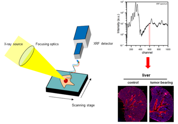
Stelling MP, Soares MA, Cardoso SC, Motta JM, Abreu J C, Antunes M J M, Freitas VG, Moraes J A, Castelo-Branco, MTL, Pérez CA, Pavão MSG. Manganese systemic distribution is modulated in vivo during tumor progression and affects tumor cell migration and invasion in vitro. Sci Rep 11, 15833, 2021. https://doi.org/10.1038/s41598-021-95190-5.
The development of photonic sources has driven various technological sectors, ranging from medicine—where advanced imaging and therapeutic techniques, such as theranostics, are employed—to agriculture, with the use of optical markers to understand plant nutrition processes. In this context, luminescent materials play a crucial role in the advancement of optoelectronic devices, data transmission and storage, lighting sources, radiation detectors, imaging systems, and light-emitting nanoparticles that can be functionalized for specific applications.
At the Carnaúba beamline, in addition to a multi-technique approach, optical processes stimulated by X-rays are investigated using XEOL combined with an X-ray nanoprobe, featuring submicron- and nanometer-scale beam sizes. This combination enables the exploration of aspects such as chemical speciation, radiation hardness, and the spatial distribution of emitting centers, with high spectral and spatial resolution. Furthermore, it opens new opportunities for the development and study of advanced photonic sources applicable to X-ray imaging devices, solar cells, energy storage materials, and more. The technique has also been employed in the investigation of natural and historical materials, aiding in the identification of the composition and chemical state of optically active centers.

TEIXEIRA, VERÔNICA C.; SILVA, JOELSON C. ; SILVA, FRANCISCO C.M. ; SZOSTAK, RODRIGO ; GUAITA, MARIA GABRIELLA D. ; KOFUKUDA, LEONARDO M. ; PICCINO NETO, ANTONIO C. ; SOTERO, ANNA P.S. ; NECKEL, ITAMAR T. ; PÉREZ, CARLOS A. ; GALANTE, DOUGLAS ; TOLENTINO, HÉLIO C.N. . X-ray excited optical luminescence at Carnaúba, the Sirius X-ray nanoprobe beamline. Optical Materials: X, v. 20, p. 100278, 2023.
KANG, MIKYUNG ; QUINTANA, JEREMY ; HU, HUIYU ; TEIXEIRA, VERÔNICA C. ; OLBERG, SVEN ; BANLA, LEOU ISMAEL ; RODRIGUEZ, VICTORIA ; HWANG, WILLIAM L. ; SCHUEMANN, JAN ; PARANGI, SAREH ; WEISSLEDER, RALPH ; MILLER, MILES A. . Sustained and Localized Drug Depot Release using Radiation¿Activated Scintillating Nanoparticles. ADVANCED MATERIALS, v. 231232, p. 1-15, 2024.
MACHADO, RAPHAEL C.L. ; FONSECA, KARINA T. ; TEIXEIRA, VERÔNICA C. ; CATALANI, LUIZ HENRIQUE ; RODRIGUES, LUCAS C.V. . Development of a Red Persistent Luminescent Composite: Electrospun Nanofiber Polymer Coating Prevents Emission Quenching by Water. Materials Today Communications, v. 35, p. 105965-9, 2023.
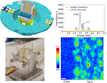
It is well known that microfluidics is a rapidly growing field attracting billions of dollars in investment every year in several strategic areas like medicine, materials science, biology, and chemistry, in which the understanding and development of new materials and/or complex systems is fundamental. Following the constant evolution of microscopy techniques, which include nowadays synchrotron-based techniques that also might explore the highly coherent beams in fourth-generation synchrotrons, we developed a microfluidic device to investigate structural, electronic and optical properties of materials at multiple scales [1] in situ/operando. It is a polyester/glass-sealed microfluidic device well-suited to combine with analytical X-ray techniques. Standard redox reactions probed the device’s throughput under electrochemical measurements, specifically cyclic voltammetry. Lastly, the 2D nanofluorescence experiments conducted at the Carnaúba beamline on pristine and silver-modified gold electrodes revealed a device highly transparent to the X-rays as designed and ready to be employed in experiments involving a liquid sample environment. Overall, the device exhibits exquisite chemical resistance to organic solvents, and its efficiency in the presence of biological samples (proteins) is remarkable.
Neckel, I.T., de Castro, L.F., Callefo, F. et al. Development of a sticker sealed microfluidic device for in situ analytical measurements using synchrotron radiation. Sci Rep 11, 23671 (2021). https://doi.org/10.1038/s41598-021-02928-2.
The study of the record of life on Earth, from its beginnings to the evolution to more complex forms that we know today, can be extensively investigated at its ultrastructural levels. With the application of the available techniques in the Carnauba beamline, by using from micro to nanometric focus, structures previously not reached by the resolution limit (such as microfossils, biominerals and other potentially preserved biotic characters) can now be explored with a high level of compositional, structural and morphological details. Paleontological materials (or materials whose biogenicity is to be investigated) can be explored with a multi-technical and multi-scale approach combining the techniques available on the beamline. Examples are the application of XRD for mineralogy determination; XANES for evaluating the oxidation states of the elements; compositional analysis by XRF (punctual spectra or mapping); XEOL to obtain luminescence spectra and pticography for 2D and 3D imaging. Paleoenvironmental details about the fossils’ context, preservation mode (taphonomy), diagenetic details (changes that occurred during the fossil’s history) as well as information regarding the biogenicity of the material can be revealed. With this, Paleontology is elevated to a science to be explored at the nanoscale.
Callefo F, Ricardi-Branco F, Alves Forancelli Pacheco ML, Cardoso AR, Noffke N, de Carvalho Teixeira V, Neckel IT, Maldanis L, Bullock E, Bower D, Moreira Silva A, Ferreira Sanchez D, Rodrigues F and Galante D (2022) Evidence for metabolic diversity in Meso-Neoproterozoic stromatolites (Vazante Group, Brazil). Front. Earth Sci. 10:804194. doi: 10.3389/feart.2022.804194
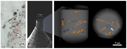
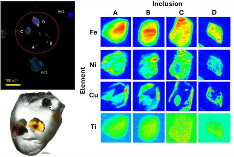
Diamonds are like time capsules that can preserve the history of our planet’s evolution. During crystallization, they encapsulate mineral inclusions originating from the deep Earth’s mantle. By analyzing the chemical composition and structure of these minerals, we can gain valuable insights into the geochemical and geophysical processes of the deep Earth, enhancing our understanding of our planet’s evolution. At the Carnaúba beamline, Tarumã station, advanced techniques such as nano-X-ray fluorescence (n-XRF) mapping and nano-X-ray tomography enable detailed analysis of the chemical composition and morphology of these inclusions. Additionally, nano-X-ray diffraction can be used to identify the mineral phases with precision. Furthermore, micro-X-ray absorption near-edge structure (μ-XANES) spectroscopy mapping at the Fe K-edge provides insights into the state of oxidation and coordination environment of iron, offering a comprehensive view of its characteristics.
The publications related to the Carnauba beamline can be found on Google Scholar’s CARNAUBA beamline.
https://scholar.google.com.br/citations?hl=pt-BR&user=FC4tp5QAAAAJ&view_op=list_works