CONTACT & STAFF
Facility E-mail: paineira@lnls.br
Coordination: Cristiane B. Rodella
Tel.: +55 19 3512 1040
E-mail: cristiane.rodella@lnls.br
Click here for more information on this Facility team.


Paineira beamline is dedicated to X-ray diffraction studies of polycrystalline materials (PXRD) housed within Sirius at the Brazilian Synchrotron Light Laboratory (LNLS). This facility will offer PXRD data collections in high resolution mode, with angular resolution estimated at 0.008º FWHM. In addition, the beamline also provides in situ and operando measurements for kinetic experiments with rapid 2D detectors, covering large angular ranges. The whole PXRD data is estimated to be acquired in seconds. The beamline hosts numerous states of the art automation processes to aid users to rapidly collect high quality data. This includes automated sample exchanges using robotics, multiple sample environments (samples holders for various types of samples, various reaction cells, devices, and accessories) and sample environments covering in-situ and operando experiments for functional materials.
The beamline is temporarily operating with a wiggler from the UVX as the X-ray source, meanwhile an undulator is in fabrication. The undulator will provide the X-ray source with a photon flux of 1013 ph/s/0.1% bw/100 mA (@15 keV) ensuring the collection of rapid, high quality diffraction patterns. It will be delivered and installed in mid 2024.
The beamline energy is fixed at 19.5keV until the commissioning of the monochromator is finished late in 2023. Then, the beamline will operate in the energy range of 5 to 30 keV (2.48 – 0.41Å) allowing for the transmission geometry experiments of high Z elements or to allow for anomalous diffraction. The flexibility of the beamline also allows for easy tuning of the photon energy to avoid fluorescence from absorption edges of chemical elements with the sample to increase data quality.
Paineira uses a Newport Heavy-Duty 3-circle diffractometer operating in Debye-Scherrer geometry (transmission or capillary geometry). Two sets of detectors are available on the diffractometer. For high resolution experiments, a set of Multi-Analyzer Crystals (MAC) will be employed. For rapid data acquisitions, a 2D arc-shaped detector (PiMega 450D).
Debye-Scherrer geometry is ideal for finely ground powder samples housed within capillaries. However, polycrystalline pellets and films may also be analyzed using the wide variety of sample holders available to users. The beamline will cover a variety of experimental cases covering fields such as, but not limited to, materials science, pharmaceuticals, catalysis, devices for energy storage and capture, such as batteries and supercapacitors, multiferroic ceramics, and environmental sciences (geoscience).
Facility E-mail: paineira@lnls.br
Coordination: Cristiane B. Rodella
Tel.: +55 19 3512 1040
E-mail: cristiane.rodella@lnls.br
Click here for more information on this Facility team.
EXP01: FAST DETECTION XPD in TRANSMISSION with SPINNER holder
Synchrotron radiation X-ray diffraction of polycrystalline powder samples (SR-PXRD) at room temperature will be performed using the fast detector (PIMEGA 450D). The detector’s resolution is 0.05° at the Full Width at Half Maximum (FWHM), and the acquisition time is about 2 minutes per sample, with a 2θ range of 1° to 108°. Samples will be spun at 50 Hz during data collection. They will be loaded into Kapton capillaries, preferably 1.0 mm in diameter, for ease of preparation (see figure 1). Depending on the transmitted signal, capillaries of 0.7 mm, 0.5 mm, 0.3 mm, 1.3 mm, and 1.57 mm are also available. The beamline energy ranges from 12 keV to 25 keV. The vertical beam size is 0.4 mm, while the horizontal is 2.0mm. You can check the transmitted signal of the sample as a function of capillary diameter and beamline energy on this website: https://paineira-xrd-tools.streamlit.app/X-ray_Attenuation_Calculator. A transmission signal of approximately 3% – 30% is required to obtain high-quality XRD data. For very absorbent samples with transmitted signals below 3%, the sample must be diluted with amorphous activated carbon or quartz supplied by the beamline team.
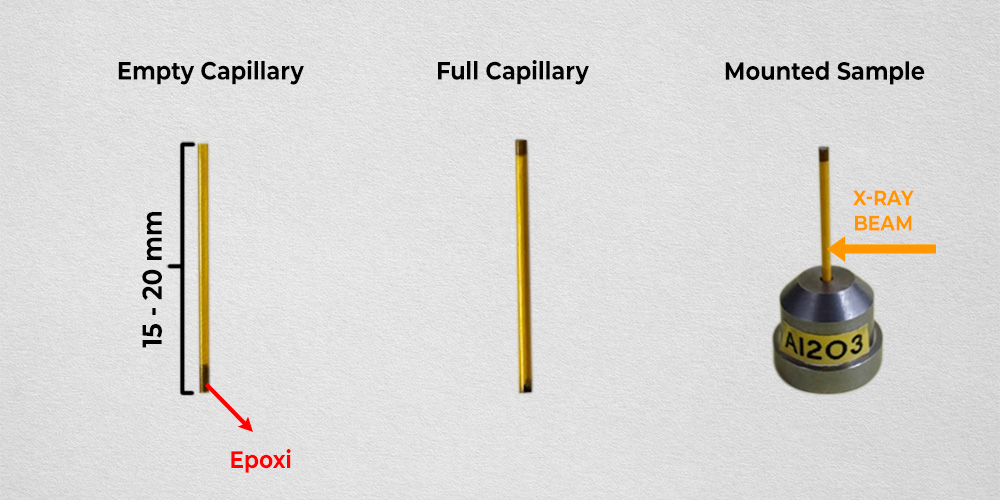
Figura 1. Etapas da montagem do capilar de Kapton para medidas de SR-PXRD com suporte giratório para análise à temperatura ambiente ou com variação de temperatura por cryojet.
EXP02: HIGH-RESOLUTION XPD in TRANSMISSION with SPINNER holder
Uses the same experimental conditions and sample preparation details as EXP01, but with the high-resolution detector (MAC) (Figure 1). The angular range available ranges from 3° to 120°, according to the sample’s characteristics. The resolution is 0.005° at the FWHM at 16 keV, and the acquisition time ranges from 2 to 4 hours, depending on the 2θ range. The beam size will be the diameter of the capillary in the vertical direction and 3.0 mm in the horizontal direction. An example of the beamtime needed to acquire a SR-PXRD pattern for Rietveld Refinement includes an angular step of 0.001°, a counting time of 0.3 seconds, and a 2θ range up to 50°. This measurement should take around 5h to finish. The staff will suggest measurements with a Q range around 7 Å-1 to optimize the 2 theta range related to beamline energy.
EXP03: IN SITU in TRANSMISSION with SPINNER holder with CRYOJET
Uses the same experimental conditions and sample preparation as EXP01 and EXP02, with both detectors, PIMEGA 450D or MAC (fast-detection and high-resolution detector, respectively). Additionally, SR-PXRD data acquisition can be performed while varying the temperature. The heat source is a cryojet, with temperature variation from 90 K to 480 K. Continuous heating and data acquisition with the fast detector, as well as temperature steps and data acquisition with the high-resolution detector, are supported. For temperature steps, it takes 10 minutes for the sample temperature to stabilize before SR-PXRD pattern acquisition begins. It is worth noting that the cryojet setpoint temperature and the temperature at the sample does not follow a strictly defined heating rate; however, it is possible to adjust the heating or cooling rate (6 K/min to 1 K/min) and set the target temperature according to the calibration curve measured offline with a thermocouple at the sample position (the most similar condition to the online condition).
EXP04-06: IN-SITU XPD in TRANSMISSION under FLUID FLOW and TEMPERATURE
Uses a capillary cell where the sample is placed in a quartz capillary between two layers of quartz wool to hold the sample (Figure 2). Alternatively, if the applied temperature is below 500 K, a Kapton capillary can be used. Both detectors can be used, but preferentially, the PIMEGA detector is applied while the temperature varies. In contrast, during isothermal treatments or at room temperature, the high-resolution detector can be used. The heat source is a hot air blower, which provides a temperature variation at the sample position from ambient up to 1148 K and the heating rate can vary from 1 to 5 K/min. The fluid can be in the gas and/or vapor phase (EXP04) and the liquid phase (EXP06), all at controlled flow. For the vapor phase, a controlled helium flow carries the fluid, and the liquid is placed in a saturator inside a thermal bath. The thermal bath temperature can vary from room temperature up to 333 K. For liquids, a peristaltic pump provides continuous, controlled flow. If a gas phase is required, you can use five different gases for the experiment. For more information on the types of gases, see Experimental conditions available to Cycle 8.
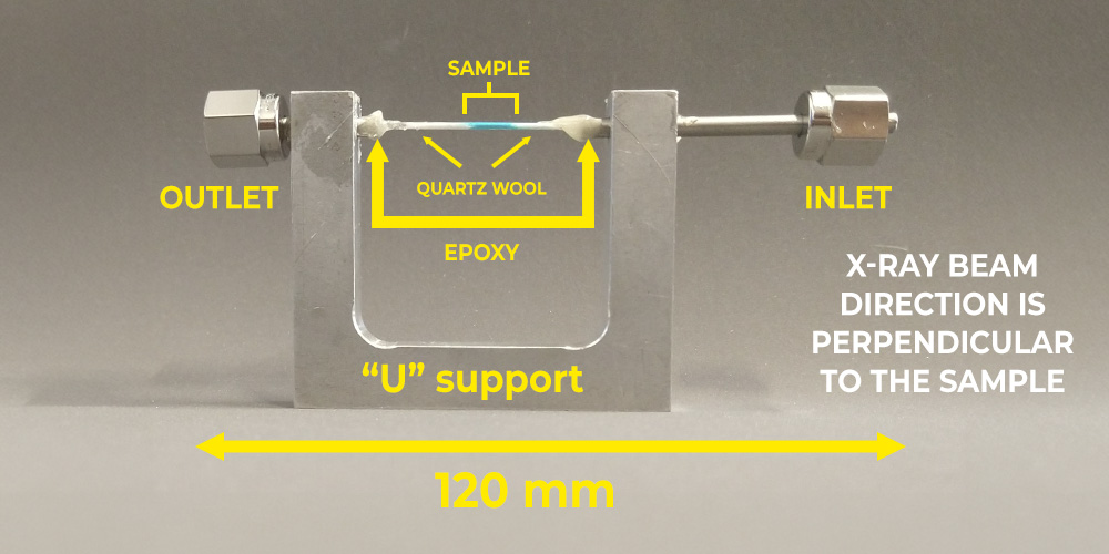
Figura 2. Montagem do reator de célula capilar para medidas de SR-PXRD in situ com fluxo de fluido e variação de temperatura.
EXP07: XPD in TRANSMISSION with FLAT PLATE holder
The flat-plate sample rotation system of Paineira beamline operates in transmission geometry and with automatic sample exchange (high-throughput) operation. During high-throughput measurements, all samples are measured under the same experimental conditions and with both detectors (PIMEGA or MAC). Flat bulk samples with parallel faces are recommended (Figure 3). The maximum sample diameter (or lateral size) is 10 mm. The flat face of the sample is fixed on the PIN with carbon tape, which is amorphous and almost transparent to X-rays. The magnetic pin has a 3.5 mm hole to allow the X-ray beam to impinge on the sample. The beam direction comes from the base to the top of the pin. The optimal sample thickness depends on the sample composition and the X-ray energy and should be calculated and adjusted by thinning before coming to the beamtime: https://paineira-xrd-tools.streamlit.app/X-ray_Attenuation_Calculator
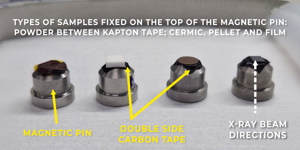
Figura 3. Exemplos de montagem de amostras para medidas de SR-PXRD em pastilhas/filmes.
EXP09: MAIL IN in TRANSMISSION with SPINNER holder
It is equal to EXP3, and sample handling will be handled by the courier system. Mail-in enables fast access to high-quality SR-PXRD measurements of crystalline powder samples via continuous proposal submission, with evaluation handled by the beamline team. For more detailed information, access Mail-in operational mode.
If you have questions, please contact the beamline team: paineira@lnls.br
Beamline Parameters and Operating Modes
The energy can vary from 12 keV to 30 keV (wavelengths from 1.03320 Å to 0.41328 Å) to the MAC detector and from 12kev to 25kev (wavelengths from 1.03320 Å to 0.49594 Å) to the PIMEGA detector. The beam size used with the PIMEGA detector is generally 0.4 mm vertically and 2.0 mm horizontally. In the case of the MAC detector, the vertical beam dimension corresponds to the capillary size, and the horizontal dimension is 3.0 mm.
Fast detector: PIMEGA 450D, with a resolution of 0.05° at the full width at half maximum (FWHM) for all energies and a fixed 2θ range from 2° to 108°.
High-resolution detector: MAC, with a resolution of 0.008° at the FWHM at 16 keV and a variable 2θ range from 2° to 120°.
The acquisition time with the fast detector is typically 1 to 2 minutes. With the high-resolution detector, the acquisition time varies according to the 2θ range and step size but is estimated to be between 2 and 4 hours per sample. We recommend measuring a Q range up to approximately 7 Å-1 for diffractions of standards intended for Rietveld refinement.
Sample Holders
For XRD measurements at room temperature and/or under temperature variation via cryojet, samples are measured in high-throughput mode in Kapton capillaries with 50 Hz rotation during analysis (Figure 1.).
Kapton capillary inner diameters: 0.3 mm, 0.5 mm, 0.7 mm, 1.00 mm, 1.12 mm, 1.37 mm, and 1.56 mm. The choice of capillary diameter depends on the beam energy and the sample composition.
Figura 1. Etapas da montagem do capilar de Kapton para medidas de SR-PXRD com suporte giratório para análise à temperatura ambiente ou com variação de temperatura por cryojet.
For highly absorbing powder samples or bulk solid samples, a flat-plate sample holder operating in transmission mode (EVL EXP07) may be used (Figure 2). Samples (powders or solids such as pellets, metals, and ceramics) are mounted using double-sided carbon tape. The X-ray window has a diameter of 3.5 mm. The sample diameter (or lateral size) must be less than 10 mm. This setup is available only under ambient conditions, and the sample rotates at 5 Hz during experiments.
Figura 3. Exemplos de montagem de amostras para medidas de SR-PXRD em pastilhas/filmes.
For experiments at temperatures ranging from 273 K to 1148 K (EVL EXP04 and EXP06), a capillary-cell reactor operating at ambient pressure is available (Figure 3). It can be used for in situ experiments involving gas, vapor, or liquid phases. Samples are loaded into quartz capillaries (0.8 mm, 1.0 mm, or 1.2 mm inner diameter, with a wall thickness of 0.01 mm and 0.08 mm) between two layers of quartz wool.
Figura 2. Montagem do reator de célula capilar para medidas de SR-PXRD in situ com fluxo de fluido e variação de temperatura.
In Situ Experiments
The cryojet allows temperature variation from 90 K to 500 K at the sample position. The cooling/heating rate can be controlled (1 K to 6 K/min), although it is not highly precise.
The hot-air blower allows temperature variation from 273 K to 1148 K at the sample position. The heating rate is precise and can vary from 2 K/min to 5 K/min.
Available gases: He, synthetic air, 3% H₂/He, 4% CH₄/He, and 5% CO₂/He.
The gas flow rate can vary from 0.1 mL/min to 30 mL/min; however, a flow rate between 5 and 10 mL/min is recommended to maintain sample stability during the experiment (preventing powder from being blown out of the capillary).
A peristaltic pump is available for liquid flow through the capillary cells, with a controlled flow rate ranging from 0.01 mL/min to 12 mL/min.
The vapor phase is delivered to the capillary cell by a helium flow passing through a saturator containing the liquid phase. The saturator temperature can vary from room temperature (22 °C) up to 60 °C.
Water, ethanol, propanol, acetone, and glycerol are approved for vapor-phase experiments. Any other liquid must be discussed in advance with the beamline staff and the chemical safety group.
Note: High-pressure experiments will not be available during this cycle due to instrumental limitations.
If you have any questions, please contact the Paineira team: paineira@lnls.br.
DESCRIPTION OF THE MAIL-IN OPERATIONAL MODE AT THE PAINEIRA BEAMLINE
1. Mail-in process
The PAINEIRA beamline for synchrotron radiation X-ray diffraction of polycrystalline materials (SR-PXRD) at SIRIUS is introducing a new operational mode: mail-in.
Mail-in enables fast access to high-quality SR-PXRD measurements of crystalline powder samples via continuous proposal submission, with evaluation handled by the beamline team.
In this initial version, this operational mode can only receive samples from Brazil and Latin American countries, including the Caribbean.
After the proposal’s acceptance, the sample holders are shipped to the proponent’s address. The user will load the Kapton capillaries with the samples and register them via the online system (ICAT) before shipping them back to CNPEM-LNLS. A detailed, user-friendly manual will guide sample preparation and procedures for returning samples to CNPEM-LNLS. The XRD measurement will be carried out within one to three months after the samples arrive at Sirius. The SR-PXRD patterns will be automatically available at the ICAT system after measurement or emailed to the proponent.
SR-PXRD measurements can be conducted using the high-resolution detector (MAC – 0.005° at the FWHM at 16 keV, available energies from 12 keV to 30 keV) or the fast detector (PIMEGA 450D – 0.05° at the FWHM at all available energies; 12 to 25 keV) at room temperature or with temperature variation using a cryojet (temperature range from 90 K to 470 K). Acquisition time, including the dead time, varies by detection type, 4h per sample in high-resolution mode, and 5 min per sample in fast detection. A total of 6 samples can be sent for high-resolution analysis, and 40 samples for fast detection measurements at room temperature.
SR-PXRD measurements will preferably be conducted at night, on holidays, and on weekends and will be fully automated. Consequently, the user will not be able to come to the beamline or follow the analysis online.
Only non-hazardous, inert samples such as minerals, clays, ceramics, soil, pharmaceuticals, alloys, oxides, hydroxides, and metals, for example, may be arranged for mail-in, as transport will be handled by the common courier system.
See below what will not be received in mail-in mode:
Inflammables, pyrophoric, explosive samples, unstable samples, oxidants, radioactive materials, biohazards, and human-derived samples.
2. SR-PXRD measurement conditions
The user selects the diffraction parameters based on the options specified in the SAU online form. SR-PXRD measurements can be conducted using the high-resolution detector (MAC – 0.005° at the FWHM at 16 keV, available energies from 12 keV to 30 keV) or the fast detector (PIMEGA 450D – 0.05° at the FWHM at all available energies; 12 to 25 keV) at room temperature or with temperature variation using a cryojet (temperature range from 90 K to 470 K). Acquisition time varies by detection type, 4h per sample in high-resolution mode, and 5 min per sample in fast detection.
Including temperature variation in the experiment will increase the time required for data acquisition. Therefore, the number of samples and available temperatures will be limited to fit in 24 h (or 3 shifts) of experiment per proposal.
The thermal treatment may vary for each sample. The options available are specified based on the cryojet’s (heat source) temperature range, the capillary material (Kapton melts at 500 K), and the maximum beamtime available for each proposal (3 shifts or 24h).
The instrumental parameter file to be used with GSAS-II software and the XRD pattern of the LaB6 NIST sample will be sent along with the user’s XRD data via email.
3. Beamline parameters
The beamline energy or wavelength will be determined by the beamline team, considering the total sample transmission of the diffracted signal, which will be optimized to a range of 3% to 30%. The team will also consider the beamline energy available at the time of the mail-in sample analysis due to the standard operational mode, as well as the sample’s fluorescence.
4. Capillary preparation
Pure samples and Kapton capillaries with an internal diameter of 1.0 mm will be preferred for comfortable preparation, but for some samples containing heavy chemical elements, 0.7 mm, 0.5 mm, and 0.3 mm will be necessary to circumvent the higher attenuation of the total X-ray transmitted signal. (See PAINEIRA_Mail-in_USER_Manual_PXRD).
PAINEIRA is usually set up at approximately 12 keV, 16 keV, 20 keV, and 25.5 keV . If you want to check the signal attenuation of your sample, you can use this online calculator: https://paineira-xrd-tools.streamlit.app/X-ray_Attenuation_Calculator
Sometimes, even with a small capillary diameter (0.5 mm or 0.3 mm) and the highest available beamline energy (around 30 keV), the transmitted signal is insufficient. In such cases, the sample must be diluted with amorphous activated carbon or quartz. The beamline team will evaluate the sample-preparation requirements and send the appropriate capillaries. Additionally, for dilution, a carbon or quartz diluent will be sent to the user with the sample holder and a manual to guide sample preparation. Only one capillary diameter size will be sent to the user. Thus, the sample composition must be similar across all capillaries.
Furthermore, after the proposal is approved, the user can email the beamline staff (paineira@lnls.br) to request the 0.3 mm capillary to achieve the highest possible resolution at PAINEIRA, regardless of the sample composition. Be aware that sample preparation in a 0.3 mm capillary is extremely difficult. It usually takes hours to properly fill one capillary.
Samples that do not meet the preparation requirements will not be measured.
Attention! Capillaries will be disposed of immediately after measurements. If the user wishes to retrieve them, they must notify PAINEIRA’s team and retrieve their samples within 30 days of the experiment. LNLS-CNPEM will not be responsible for returning measured capillaries.
Experiments that require conditions not available in mail-in mode can be submitted via standard proposal submission during the open call.
If you have questions, please contact the beamline team: paineira@lnls.br
| Element | Type | Position [m] | Description |
|---|---|---|---|
| Source | Insertion device | Wiggler 1T (temporary source) | |
| White beam slit | Slit | 27.9 | Determination of beam divergence |
| Beam diagnostics | Diagnosis | 28.2 | Visualization and diagnosis of the white beam |
| Double crystal monochromator | Bruker Monochromator | 30.0 | Monochromatization |
| Beam diagnostics | 32.0 | Monochrome beam visualization and diagnosis | |
| Monochrome beam slit | 43.4 | Monochrome beam slit | |
| Beam diagnostics | 43.4 | Initial intensity counter (I0) | |
| X-ray attenuator | 44.8 | Beam attenuator | |
| Robotic arm | GP25 – Motoman | 45.2 | Sample room changer |
| In situ design control module | Development in house | 45.5 | Gas flow control and design control |
| Diffractometer | Heavy-Duty, 3-circles | 46.0 | Sample alignment with incident beam and diffracted beam detectors |
| Detection system | MAC – Oxford FMB | 46.0 | Set of 8 independent analyzer crystals with ~2° 2θ separation of scintillating detectors (FMB-Oxford) |
| Detection system | ARCPIX (in-house development) | 46.0 | Fast arc-shaped detector with 100° angular coverage |
| Robotic arm | GP8 – Motoman | 46.5 | Sample changer |
| Sample Magazine | Development in house | 47.0 | Sample storage carousel where GP8 robot will transfer sample |
| X-ray beam visualization | X-ray and photodiode eyes | 47.5 | Transmitted beam viewer |
| Parameter | Value | Condition |
|---|---|---|
| Energy range | 5 – 20 keV 14 – 30 keV |
Si(111) Si(311) |
| Flux at sample [ph/s] | ~1013 | 15 keV |
| Energy resolution (ΔE/E) | ~10-4 | 15 keV |
| Angular resolution (MAC) | 0.008° | FWHM @ 15 keV |
| Angular range in 2θ (MAC) | 3 – 145° | |
| Angular resolution (Pimega 450D) | 0.05° | (all energies) |
| Angular range at 2θ (Pimega 450D) | 3° – 100° | |
| Beam dimensions [mm x mm] | 1.1 (v) x 1.7 (h) | FWHM @ 15 keV |
| Beam divergence [μrad] | 25 (v) x 37 (h) | FWHM @ 15 keV |
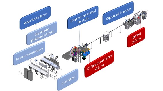
In addition to the experimental hutch, the PAINEIRA beamline has a room for sample manipulation (Figure 1), with direct access to the experimental hutch, through a second door to facilitate the arrival of samples to the diffractometer. Connected to this room is the control room of the beamline, where the user operates the beamline, processes the acquired data, and visualizes all the experimental parameters of the experiment. Finally, the beamline has an instrumentation room.
The experimental station has been designed to optimize the amount of synchrotron light users have access to during their beamtime. To provide fast data acquisition with various experimental setups, the diffractometer (Figure 2.1), uses two sets of fixed detectors (Fig. 2.2 and Fig. 2.3) with automated sample exchange via a sample magazine (Fig. 2.4) with robotics dedicated to automated and autonomous sample exchange. (Fig. 2.5).
Robotics and automation for the operation and use of thermal blowers for experiments ( cryojet or gas blower – Fig. 2.6) will further aid in providing a rapidly exchangeable experimental platform for various types of experiments (Fig. 2.7). When the beamline is operated in high-throughput mode, the beamline becomes fully autonomous, easily controlled remotely, or left to measure and screen wide ranges of samples with ease and speed. When gas or liquid flow is required, an automated, fixed in-line control module aids the user to easily change the reaction environment to simulate real world conditions (Fig. 2.8). Finally, to study chemical reactions, mass spectrometer analyzers (Fig. 2.9) and (Fig. 2.10) micro gas chromatographs are available and connectable to the diffractometer’s reactors.
To meet the demand to conduct cutting edge research, the two types of detectors can be used consecutively and prioritized to work with the required instrumentation for ex situ experiments, as well as in situ and operando.
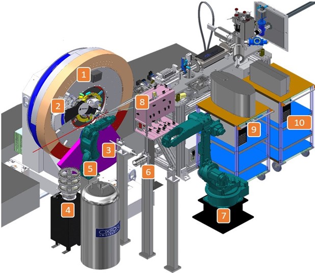
Figure 2. Design of the Paineira beamline experimental station: 1) Newport Heavy-Duty 3-circle diffractometer; 2) High resolution detector; 3) fast arc detector spanning 109° in 2 θ ; 4) Sample magazine with 320 capillary capacity; 5) Robotic arm for automated exchange; 6) Supports for the cryojet and hot air blower to be used for cooling and heating the samples; 7) Robotic arm to place the cryojet or hot air blower at the sample position during experiments; 8) Gas flow control module and in situ and operando experimental control module; 9) Mass spectrometer; and 10) Micro GC.
Paineira provides sample holders, reaction cells and analytical equipment for measurements to cover a wide range of experimental conditions. In addition, the Paineira group will continue developing new instrumentation for in situ and operando experiments for the scientific user.
The table below shows systems that have already been developed and are available at beamline in operation. It is strongly recommended to contact the beamline staff to check the availability and flexibility of the instrumentation. Moreover, new sample environment development will be welcomed.
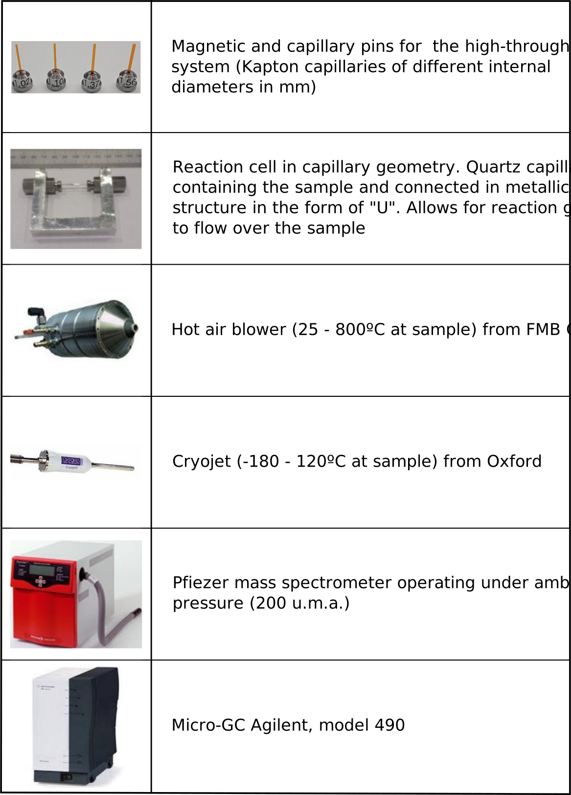
Figure 3: Sample holder, cell and accessories for X-ray diffraction experiments of polycrystals in PAINEIRA.
High-resolution mode features a system of 8 Si analyzer crystals (111), aligned in front of point scintillating detectors (Fig. 3). The X-ray beam diffracted by the sample passes through the analyzer crystals set at the appropriate Bragg angle. The angle is the Darwin width of the Si(111) crystal only allows diffracted x-rays satisfying the Bragg condition to be detected. For example, at 15 keV with an analyzing crystal of Si(111), the Darwin width is approximately 3.55 arcsec. This high-resolution setup will produce highly resolved diffraction peaks while removing background as well as florescence signals for improved signal to noise ratios. Diffraction peaks with full width at half maximum (FWHM) of the order of 0.008° at 2θ will be resolvable. However, this detection mode with point-to-point collection requires relatively longer data collection times. Scans covering a 2θ angular range from 5-100° at 15 keV will take around 1 hour to complete.
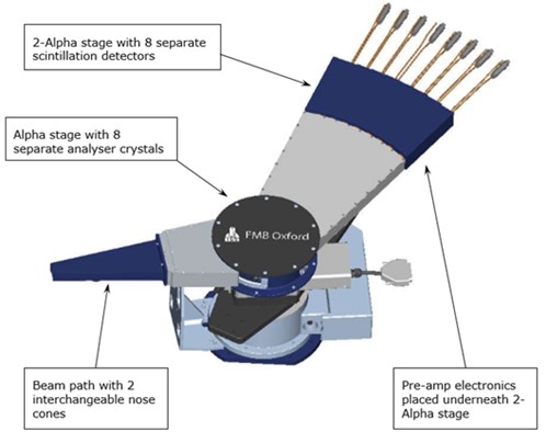
Figure 3. The FMB-Oxford MAC system for high-resolution experiments.
The fast acquisition, medium resolution detector (0.07° FWHM resolution) is an internal development of the LNLS detector group in partnership with Pi-Tecnologia. The Pimega 450D is a detection system based on Medipix3RX ASIC detectors, arranged in a 2D arc shape with wide angular coverage and installed on the delta circle of the diffractometer, as indicated in Fig. 2.3). This mode of data collection will optimize measurement time where rapid data collection is required while ensuring good data quality and resolution. The setup aims to meet the requirements of scientific cases that require rapid detection when a sample is undergoing structural transformation and required high temporal resolution (seconds/pattern) and/or has sensitivity to X-ray exposure to reduce sample damage induced by the beam.
The Pimega 450D contains 10 modules, with 2 Si elements each, installed in an arc around the delta circle of the diffractometer. Each Si element (Medipix3RX ASIC) uses a 256 x 1550 pixel chip with each pixel 55 μm x 55 μm placed 89 cm from the sample resulting in an angular resolution of 0.05° (FWHM). The read rate will be up to 1000 frames per second. The detector will cover a large angular 2θ range from 0 to 100°, with entire diffraction patterns collected in a matter of seconds. As it is a 2D detector (256 pixels x 30,060 pixels), an azimuthal integration will be performed for each measurement to transform the image of the X-ray diffraction pattern into an Intensity versus 2θ plot. Furthermore, to improve the collection statistics the detector will swivel between two positions to cover any gaps between each detector module. Thus, two individual measurements with offset of the circle delta will be added to produce a single continuous diffractogram and a software will make the superimposition and necessary corrections for the result (graph Intensity vs 2θ) to be quickly visualized by the user.
Debye-Scherrer geometry works ideally using a powdered sample inserted into capillary tubes. The available capillaries (materials and capillary diameters) are presented in Table 1. It is noteworthy that the kapton capillaries will be used for Paineira’s high-throughput operating system. Borosilicate and quartz capillaries will be used in the capillary cell.
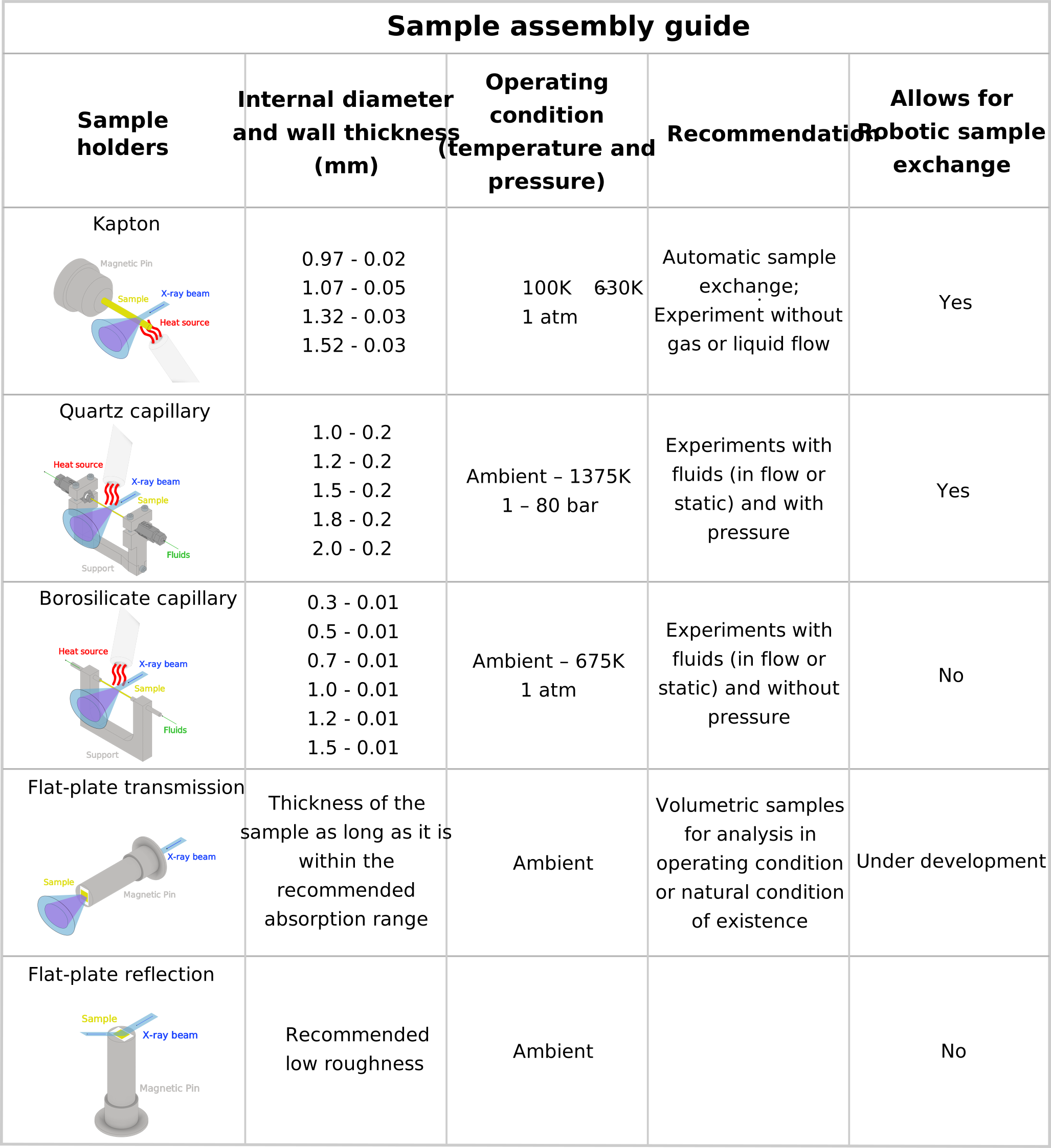
Table 1. Sample holders available in the Paineira beamline and their specifications.
Calculation of sample absorption as a function of composition, packing density (~0.6 for powder samples and ~1 for volumetric bodies), capillary diameter (or thickness of volumetric sample) and wavelength can easily be done using the following online calculator. To allow for optimized signal in transmission geometry, the X-ray absorption coefficient must be calculated to assist in selecting the energy of the experiment (19.5keV) and the thickness of the capillary (or of the sample, in the case of the flat-plate).
https://11bm.xray.aps.anl.gov/absorb/absorb.php
In cases where it is necessary to decrease the absorption coefficient, the packing density can be decreased by mixing the sample with a low-density, amorphous material. If you have any questions regarding sample preparation and experimental conditions, please do not hesitate to contact the beamline team for assistance.
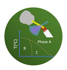
The development of new materials has contributed to the search for novel solutions to the worlds technological, environmental and health problems. In this case, to design and control physical properties of materials, it is vital to understand structure in both ambient conditions, as well as a function of temperature. Measurements such as these allow for better understanding of synthesis methods or analysis of phase transitions and construction of phase diagrams for example.
Here, X-ray diffraction with automatic sample change can be used as a function of composition to analyze the crystalline phases and distinguish between polymorphic phases at room temperature. Or, when interesting, to know the behavior of crystal structures as a function of temperature.
Paineira will make it possible to combine different angular, temporal and temperature ranges resolutions. The results of these crystal structure thermal dependence experiments reveal the behavior of the crystal structure, the existence of phase transitions, as well as the order of the transition (first or second order) and its character (displacive or order-disorder). These results, correlated with the thermal dependence of physical properties (dielectric, magnetic, thermal, elastic, optical analysis, among others) are fundamental for the control and operation of these new materials and for the proposal of new technologies.
The structural properties of functional materials, such as catalysts, change depending on the variation of reaction parameters (fluid type, temperature, and time, for example), as well as application conditions. Thus, it is of great importance that the studies of the physical and chemical properties of these materials are carried out under in-situ conditions (control of one or more reaction parameters) and, if possible, the operation (operating) should be characterized under conditions. Thus, at Paineira it will be possible to install a reaction cell, called a capillary, where the sample will be deposited. The cell will allow controlled flow of gas, liquid, or both and with ambient pressure variation up to 80 bar. It will also be possible to pass a controlled flow of steam at ambient pressure and heat the sample to 800 ºC at controlled rates and/or temperature thresholds. The references below report studies of this type that will be possible in the Sirius Paineira beamline.
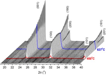
Glauco F. Leal, Dean H. Barrett, Heloise Carrer, Santiago J. A. Figueroa, Erico Teixeira-Neto, Antonio Aprigio S. Curvelo, Cristiane B. Rodella. J. Phys. Chem. C, 2019, 123, 5, 3130–3143. DOI: 10.1021/acs.jpcc.8b09177
The structural properties of electrocatalysts are directly related to the performance, lifetime and charge cycles of energy storage devices such as batteries and supercapacitors. Electrocalyzers are generally metals and/or transition metal oxides that are normally dispersed in carbon and form the cathode of these devices. Structural changes, as well as the formation of crystalline by-products during the loading and unloading processes of the devices, occur during their operation. Therefore, these make up more examples of scientific cases where PXRD measurements need to be collected during the operation of these devices. Thus, Paineira will also develop electrochemical cells and cells supports to be installed in the light line and connected to a potentiostat. Thus, allowing the detection of structural changes of polycrystalline materials from these energy storage devices, as shown in the references below:
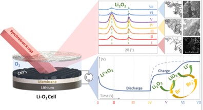
Leticia F. Cremasco, Chayene G. Anchieta, Thayane C. M. Nepel, André N. Miranda, Bianca P. Sousa, Cristiane B. Rodella, Rubens M. Filho, Gustavo Doubek. ACS Appl. Mater. Interfaces 2021, 13, 11, 13123–13131. DOI: 10.1021/acsami.0c21791
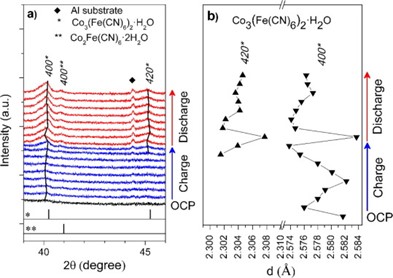
Bruno Morandi Pires, Willian Gonçalves Nunes, Bruno Guilherme Freitas, Francisca Elenice Rodrigues Oliveira, Vera Katic, Cristiane Barbieri Rodella, Leonardo Morais Da Silva, Hudson Zanin. Journal of Energy Chemistry Volume 54, March 2021, Pages 53-62. DOI: 10.1016/j.jechem.2020.05.045