CONTACT & STAFF
Facility E-mail: sabia@lnls.br
Coordination: Julio C. Cezar
Tel.: +55 19 3512 1292
E-mail: julio.cezar@lnls.br
Click here for more information on this Facility team.


The SABIÁ (Soft X-Ray ABsorption spectroscopy and ImAging) beamline operates in the soft X-ray range using a Delta-type elliptical undulator with polarization control and plane grating monochromator. The main possible analyses are X-ray absorption spectroscopy (XAS) and photoemission electron microscopy (PEEM). In addition, several variations of dichroism in X-ray absorption allow the investigation of structural and magnetic properties with chemical selectivity. This aspect benefits from the energy range of photons, which corresponds to the $L_2$ and $L_3$ absorption edges of $3d$ transition metals, such as manganese, iron, cobalt, the $K$ absorption edges of light elements such as carbon, nitrogen, and oxygen, and the $M_4$ and $M_5$ edges of rare earths. The same dichroism techniques can be applied with spatial resolution of the order of tens of nanometers using the electron photoemission microscope.
The SABIÁ beamline will work in close collaboration with the In-situ Growth Laboratory (LCIS), which has a versatile system for the preparation and pre-characterization of thin film and heterostructure samples. These samples can be transferred under ultra-high vacuum conditions for analysis in the SABIÁ line through portable vacuum chambers.
Facility E-mail: sabia@lnls.br
Coordination: Julio C. Cezar
Tel.: +55 19 3512 1292
E-mail: julio.cezar@lnls.br
Click here for more information on this Facility team.
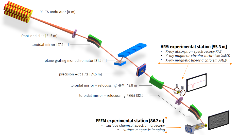
| Element | Kind | Position [m] | Description |
| SOURCE | Insertion Device | 0.0 | DELTA ondulator, 52.5 mm period, 1.2 m long. Possibility of controlling the polarization of the X-ray beam (linear or circular). |
| FOE | Slits | 21.5 | Power-absorbing slits and beam size selection. |
| M1 | Toroidal Mirror | 27.5 | Absorber of the main thermal charge coming from the ring. It focuses the ondulator beam on the exit slit. |
| PGM | Monochromator | 31.5 | Flat mirror and variable line density grating. It vertically disperses the beam as a function of the photon energy. |
| ES | Exit slits | 39.5 | Selects photons with desired energy from the beam dispersed by the PGM |
| M2 | Toroidal Mirror | 43.8 | Refocuses beam on arm A (HFM) |
| HFM | Experimental Station | 55.3 | HFM, superconductor coil, XAS, XMCD, XMLD with magnetic field and sample temperature control. |
| M3 | Toroidal Mirror | 82.5 | Refocuses beam on B arm, PEEM microscope |
| PEEM | Experimental Station | 86.7 | Photoemission electron microscope. Chemical and magnetic imaging of surfaces |
| Parameter | Value | Condition |
| Energy range [eV] | 200 – 1600 | |
| Energy resolution [ΔE/E] | ~5×10⁻⁴ | |
| Photon energy sweep | Yes | |
| Beam Size [μm2] | 20 x 80 | Magnet |
| Beam Size [μm2] | Variable, down to 15 x 15 | PEEM |
Variations in the crystal structure of materials cause X-ray absorption to differ depending on the orientation between the electric field of the X-ray beam and the crystal axes of the sample. This effect is known as X-ray linear dichroism (XLD) and is a powerful source of information about variations in the structure of the interfaces and surfaces of thin, multilayer films. In addition, the absorption of linearly polarized radiation can vary with the magnetization of the sample. In this case, we are dealing with X-ray linear magnetic dichroism (XLMD) and we have magnetic information with chemical sensitivity.
Materials that have a non-zero magnetic moment absorb differently the two possible circularly polarized X-ray helicities (left and right circular polarizations). This difference is called circular dichroism, and when performed at one of the absorption edges the components of the sample allow to obtain the magnetic contributions of each chemical element independently. In addition, in many cases it is possible to determine the spin and orbital components of magnetism. The dichroism sign is usually maximum at the peak edge and absorption of each element. By keeping the energy fixed at this point in the spectrum and varying the applied magnetic field, it is possible to obtain magnetic hysteresis curves for each element in the composition of the sample. This aspect is particularly important in the characterization of new permanent magnets.
Matter interacting with the X-ray beam emits electrons. This effect is particularly important in the region of soft X-rays. Using a column similar to that of a transmission electron microscope, it is possible to obtain images based on the electrons emitted in the absorption process. This is the basic principle of the PEEM (photoemission electron microscopy) technique, which makes possible to obtain spectroscopic information with a spatial resolution of up to a few tens of nanometers. In addition, the XMLD and XMCD methods are still valid, and by properly using the polarization of the X-ray beam, it is possible to obtain magnetic information with such spatial resolution. This possibility is particularly interesting in the study of magnetic domain walls and their dynamics. In addition, given the inherent chemical sensitivity to X-ray absorption, the technique has great potential in geosciences, environmental studies, among others, where it is important to locate the spatial concentrations of the various chemical elements in the sample.