CONTACT & STAFF
Facility E-mail: ipe@lnls.br
Coordination: Tulio C. R. Rocha
Tel.: +55 19 3512 1292
E-mail: tulio.rocha@lnls.br
Click here for more information on this Facility team.


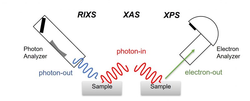
IPÊ1 beamline is dedicated to high resolution resonant Inelastic X-ray scattering (RIXS), X-ray absorption spectroscopy (XAS) and X-ray photoelectron spectroscopy (XPS) in the soft X-ray range (100 – 2000 eV). These complimentary techniques of spectroscopy and scattering allow characterization of the chemical composition, electronic structure, and elementary excitations in solids and molecular systems.
Analysis of XAS and XPS using soft X-ray allow quantitative determination of chemical composition of the near surface region of solid samples, which is a crucial information to several areas from thin films and electronic devices to enzymes and catalysis. XPS and XAS are also important to determine the electronic structure of materials. RIXS goes one step further in the understanding of the properties of materials by directly probing the spectrum and dispersion of excitations, such as excitons, magnons, and phonons, which determine several materials’ properties, such as magnetism, electronic conductivity, thermal conductivity, polarizability, among others.
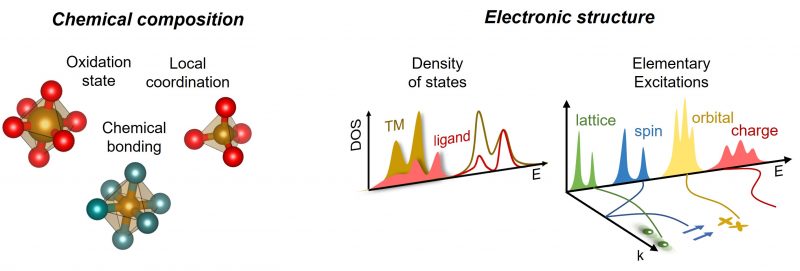
This broad view of the electronic structure and excitations provided by IPÊ might assist not only the understanding of materials properties and but also guide the design of new materials with tailored functionalities. Also, it might be used to fine tune theoretical models widely used to describe the electronic structure of solids and molecules.
1 Inelastic scattering and PhotoElectron spectroscopy
Facility E-mail: ipe@lnls.br
Coordination: Tulio C. R. Rocha
Tel.: +55 19 3512 1292
E-mail: tulio.rocha@lnls.br
Click here for more information on this Facility team.
The optics of IPÊ deflect the X-ray beam into two branches allowing the installation of two endstations permanently connected to the beamline. One of these endstations is dedicated to RIXS and the other to XPS, while both can perform XAS measurements.
Important: proposals with standard XPS measurements aiming to obtain information about the surface composition and chemical speciation should not be executed at IPE, but forward to LNNano (information here) or performed elsewhere.
XPS is a surface-sensitive quantitative spectroscopic technique based on the photoelectric effect that can identify the elements that exist within a material (elemental composition) or are covering its surface (1-10 nm). XPS is a powerful measurement technique because it not only shows what elements are present but also what other elements they are bonded to. XPS and XAS are also important to determine the electronic structure of materials. While XPS probes the occupied states of the core and valence electrons, XAS measure the unoccupied states projected on the absorbing atom.
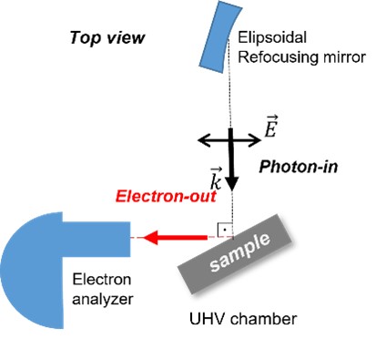
Geometry of the experiments at the XPS endstation of IPE.
XPS measurements should be performed on the IPE beamline only in special cases, where proponents clearly demonstrate the need for synchrotron radiation (ideally presenting standard XPS as previous characterization in the proposal). Examples of cases that justify the use of synchrotron light include species with concentrations below the detection limit or chemical shifts smaller than the resolution of standard equipments. Other photoemission experiments that justify the use of the beamline include chemical mapping that exploits the micrometric beamsize, valence band using low-energy photons, resonant photoemission, and resonant Auger electron spectroscopy. Additionally, experiments with complex sample environments that are unfeasible with commercial instruments, such as cryostats, in situ heating, strain cells, devices with applied electrical potential, liquid microjets, among others, also justify the use of the IPE beamline.
Regarding experiments to measure depth profiles using variable photon energy, a careful evaluation is essential. In the specific case of samples with smooth surfaces, these experiments justify the use of the beamline only in situations where sample rotation is challenging with conventional equipment, preventing depth profile measurement by varying the angle. This is the case, for example, of micrometric samples or experiments in complex sample environments.
The XPS endstation is composed of an electron analyzer SPECS Phoibos 150 with a 9-channeltron detector connected to a main chamber, which has a manipulator with 4-axis (X, Y, Z, theta) that performs positioning and rotation of the samples in the X-ray beam. A pre-chamber serves both as a load lock for fast sample loading and a reactor for heat treatments under a controlled gas atmosphere. The electron analyzer is positioned in the horizontal plane at a fixed angle of 90o relative to the incident X-ray beam, but the sample can be rotated to adjust the desired incidence angle (θin) and photoemission angle (θout). Several experiments are possible at the XPS endstation.
| Experiment | Information | STATUS |
| Core-level XPS | Surface composition and speciation | Available |
| Depth profile | Depth distribution of species | Available |
| XAS in TEY mode | Near surface local chemical environment | Available |
| XAS in FY mode | Bulk sensitive XAS | Available |
| XAS in AEY mode | Surface sensitive XAS | Available |
| HV annealing | Ex situ heating in high vacuum | Available |
| RAES | Core-hole lifetime and dynamics | Available |
| RPES | Projected DOS | Available |
| Valence UPS | Electronic structure, band bending | Available |
| Sputtering | In situ surface cleaning | Available |
| Chemical mapping | lateral distribution of species | Commissioning |
| Gas annealing | Ex situ heating in gas atmosphere | Development |
| Vacuum suitcase | Sample transfer under UHV from LCIS | Development |
Samples for XPS can be solids in any form (powders, foils, thin films, composites, or crystals), but must be compatible with ultra-high vacuum (UHV) conditions. The main chamber normally operates at 1-2 10-8 mbar, but if the experiment requires, it can be baked-out down to ~ 8 10 -10 mbar (contact staff). Be aware that samples with considerable degassing (wet or high porosity) must be kept longer at the load lock until safe pressure for transfer is reached. Also, the samples must be moderately conductive to avoid surface charging, which distorts the XPS measurements. In case of insulating samples, a flood gun is available to electrically neutralize the surface minimizing shifts and lineshape distortions.
Samples are typically fixed on metal plates that are attached to sample holder and transferred under UHV to the manipulator. We provide two model of sample holders (~30×30 mm2) that can accommodate multiple samples and special holders for annealing. Detailed drawings of the standard sample plates in pdf format can be found here. Custom sample environments could be used if required by the experiment, but this must be discussed with the beamline staff in advance. The beamline has a small sample preparation room with common supplies, tools and equipment used for the manipulation of solids. Contact staff if your sample require special preparation conditions.
XPS experiments in the presence of gases or liquids are NOT possible with the current setup.
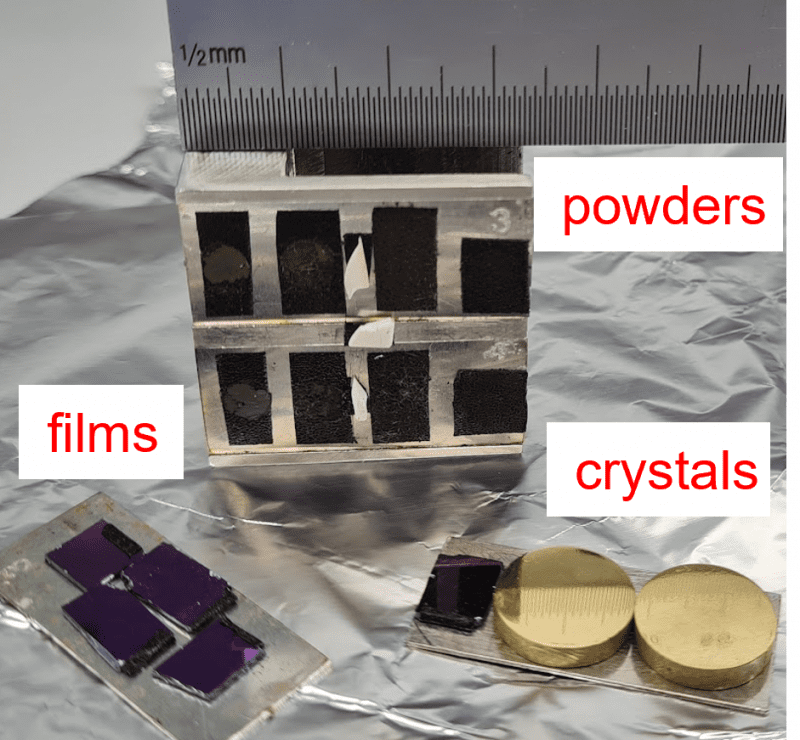
Standard XPS sample holder for solid samples fixed with carbon tape on metallic plates.
Please do not hesitate to contact the beamline staff for more information on samples and environments.
Resonant inelastic X-ray scattering is a photon-in/photon-out technique that measures the energy and momentum change of photons scattered by the sample. The energy and momentum lost in the inelastic process are transferred to intrinsic excitations inside the material under study. RIXS has some unique features that distinguish it from other inelastic techniques based on the scattering of neutrons (INS), electrons (EELS), or light (Raman), commonly used to study elementary excitations. It is bulk sensitive, requires only small sample volumes, it is element and orbital specific, polarization dependent, and covers a large scattering phase-space. With soft X rays in the range of 200 to 1200 eV, RIXS is particularly useful for studying 3d transition metals and light elements in complex oxides, correlated electron materials, molecular complexes, catalysts, and metalloenzymes, among others.
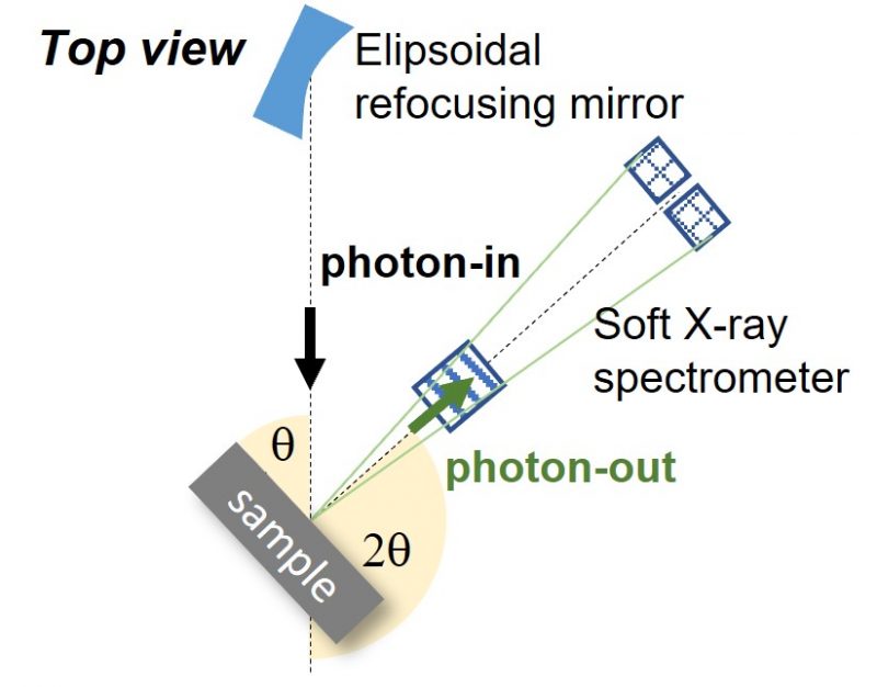
Geometry of the experiments at the RIXS endstation of IPE.
The RIXS endstation is composed by an in–house developed soft X-ray spectrometer based on a VLS cylindrical diffraction grating (2000 l/mm) and a high spatial resolution area detector (EMCCD RIXSCAM2). The spectrometer is connected to a main chamber with differentially pumped rotatable flange that allows continuous rotation under vacuum. The radiation scattered by the sample is collected in the horizontal plane at a variable 2q angle of 30-150 deg relative to the incident X-rays. They are dispersed by a cylindrical VLS grating into a set of 2 EMCCD detectors that performs single photon counting. A high precision manipulator with 4 axis (X, Y, Z, theta) performs positioning of the samples in the X-ray beam and rotation to control the momentum transfer direction relative to the sample surface.
Some relevant informations on the current status can be found in the following table:
| Experiment | Information | STATUS |
| Current res. power | ΔE/E ~ 8000 | Available |
| Cryo stage | LHe cooling down to < 10 K | Available |
| Liquids | Si3N4 membrane cell | Development |
| Single crystal | In situ rotation chi-phi | Development |
| Target res. power | ΔE/E > 30000 with full length undulator | Development |
Samples for RIXS can be solids in any form (powders, foils, thin films, composites, or crystals), but must be compatible with UHV conditions. However, high quality single crystals or epitaxial thin films are necessary for high resolution or momentum resolved measurements. We typically fix the samples on carrier plates that are transferred under UHV to the sample holder in the manipulator. Our standard sample carriers are flag style metal plates, compatible with SPECS sample holders. The main chamber normally operates at a base pressure of 5 10-9 mbar. It cannot be baked because of the special sealing for continuous rotations. The beamline provides a dedicated room for sample preparation with common supplies, tools and equipment used for the manipulation of solids. Contact staff if your sample require special preparation conditions.
Sample environment inserts with different functionalities can be mounted on the manipulator, but it must be specified in the proposal and discussed with the beamline staff in advance. Currently, we have a standard room temperature insert that can hold up to 3 sample plates, a cryogenic insert (ColdEdge open cycle cryostat) that can hold only 1 sample plate. The standard operating temperature range is 25 – 400 K with LHe and 75 – 400 K with LN2. With the insertion of a thermal shield, it is possible to achieve < 10 K with LHe. Additionally, we are working on a static cell for liquids based on Si3N4 membranes and a cryogenic insert with in-vacuum phi-chi rotations for single crystal alignment.
Please do not hesitate to contact the beamline staff for more information on samples and environments.
The optics of IPÊ was specifically designed to exploit the fully diffraction limited soft X-ray source at the Sirius storage ring delivering very high resolving power and small spot size required for high-resolution RIXS experiments.
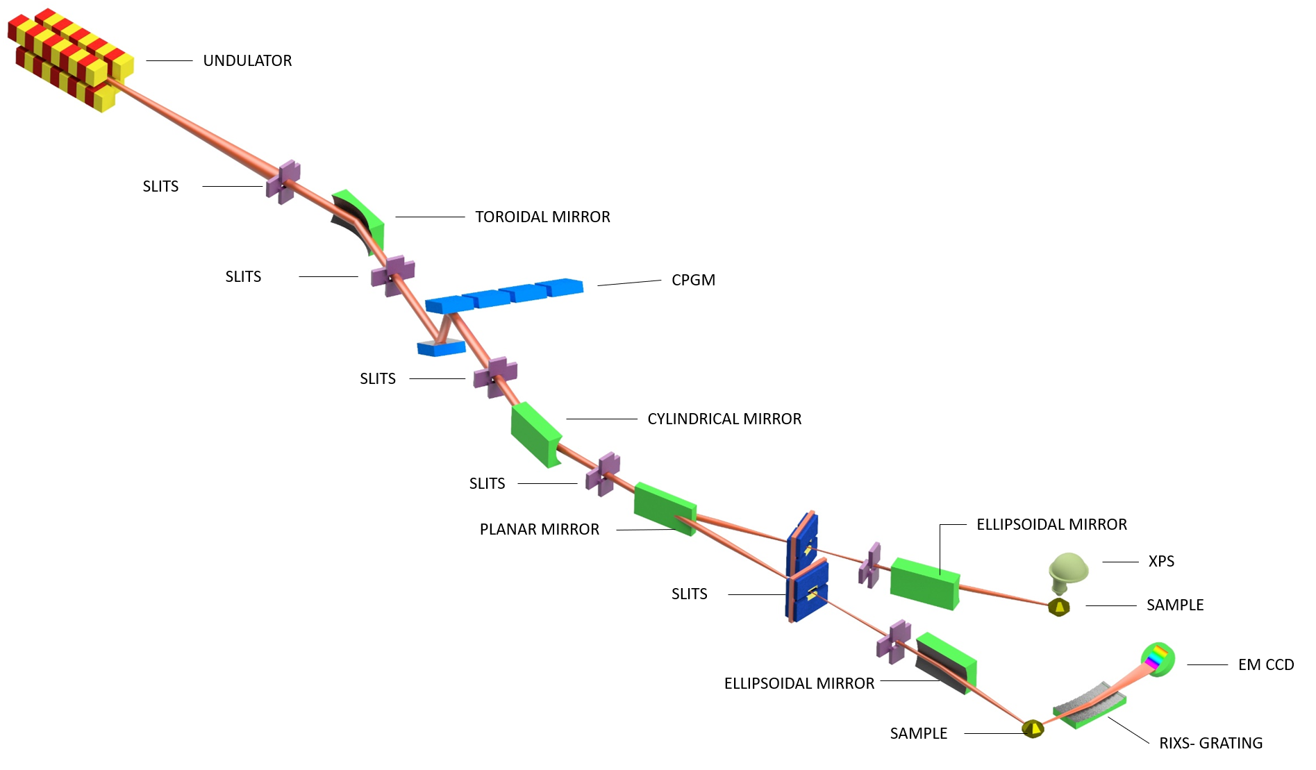
| Element | Type | Position [m] | Description |
| Source | Insertion device | 0 | Planar Undulator (1.2 m/58 mm) |
| M1 | Toroidal mirror | 27 | Horizontal focusing and vertical collimation |
| M2 | Plane mirror | 28.5 | Change grating incidence angle |
| M3 | Diffraction gratings | 29 | Energy dispersion (1100/400 l/mm) |
| M4 | Cylindrical mirror | 31 | Vertical focusing at the slit |
| M5 | Plane mirror | 33 | Branch selection |
| M6 | Ellipsoidal mirror | 81 | Microfocus at XPS endstation |
| M7 | Ellipsoidal mirror | 91.5 | Microfocos at RIXS endstation |
| M8 | RIXS grating | 93 | Energy dispersion (2000 l/mm) |
| Parameter | Value | Condition |
| Energy range | 100 -2000 eV | Horizontal polarization |
| Resolving power | 60.000 | 930 eV, cff = 5 |
| Spot size RIXS | 0.8 x 3.4 μm | 930 eV, cff=5 |
| Spot size XPS | 4 x 5 μm | 930 eV, cff = 2.2 |