CONTACT & STAFF
Facility Tel.: +55 19 3517 5157
Facility E-mail: imbuia@lnls.br
Coordination: Raul de O. Freitas
Tel.: +55 19 3517 5060
E-mail: raul.freitas@lnls.br
Click here for more information on this Facility team.


IMBUIA1 is the infrared (IR) beamline of the Brazilian Synchrotron Light Laboratory (LNLS) in operation at the Sirius storage ring. The beamline is a state-of-art resource comprising an experimental station dedicated to nanoscale synchrotron IR spectral-imaging (IMBUIA-nano) covering the entire mid-IR range. The design goal for IMBUIA was to provide a user-friendly, multidisciplinary and highly sensitive analytical tool benefited by the enhanced stability of a 4thgeneration storage ring.
The synchrotron IR extraction port (~5×5 mrad2) delivers a photon flux of ~3×10¹² ph/s/0.1%bw/100 mA at 10 µm wavelength. Energy coverage is from 105 meV to 520 meV (12 – 2.4 µm) allowing analyses of organic and inorganic materials in general. Custom sample environments can be designed for specific experiments.
1Infrared Multiscale Beamline for Ultra-resolved Imaging Applications
Facility Tel.: +55 19 3517 5157
Facility E-mail: imbuia@lnls.br
Coordination: Raul de O. Freitas
Tel.: +55 19 3517 5060
E-mail: raul.freitas@lnls.br
Click here for more information on this Facility team.

| ELEMENT | TYPE | POSITION [m] | DESRIPTION |
|---|---|---|---|
| Source | Bending Magnet | 0.00 | 0.56 T B2 dipole |
| M1 | Water cooled flat mirror | 1.20 | Au/Glidcop mirror |
| W1 | Optical window | 1.62 | CVD diamond |
| M2 | Flat mirror | 3.79 | Au/Al mirror |
| M3 | Flat mirror | 4.16 | Au/Al mirror |
| W2 | Optical window | 4.93 | BaF2 |
| M5 | Elliptical mirror | 5.20 | Au/Al mirror |
| 2nd source | Focal point | 5.60 | – |
| M6 | Parabolic mirror | 6.04 | Au/Al mirror |
| M4 | Parabolic mirror | 4.25 | Au/Al mirror |
| W3 | Optical window | 14.20 | BaF2 |
| PARAMETER | VALUE | CONDITION |
|---|---|---|
| Energy Range [meV] | 105 – 521 | MCT detector + synchrotron |
| Wavelength Range [μm] | 11.8 – 2.4 | MCT detector + synchrotron |
| Frequency Range [cm-1] | 850 – 4200 | MCT detector + synchrotron |
| Spectral Resolution [cm-1] | up to 1 up to 0.1 |
s-SNOM, SINS micro-FTIR |
| Energy Selection | FT Processing FT Processing Source tuning |
SINS micro-FTIR s-SNOM (laser QCL) |
| Probe dimension [μm] | 0.025 ~3 (diffraction limited) |
s-SNOM, SINS micro-FTIR |
IMBUIA is an analytical complex that comprises two experimental stations dedicated to synchrotron infrared micro- and nano-spectral imaging. The IMBUIA-nano station operates an ultra-microscopy setup based on scattering Scanning Near-field Optical Microscopy (s-SNOM) combined with IR synchrotron for continuous coverage across the mid- to far-IR spectral range. Additional table-top sources, such as Quantum Cascade (QCL), allow for mid-IR narrowband nano-imaging.

Overview of the IMBUIA experimental stations, control rooms and instrument lab space.
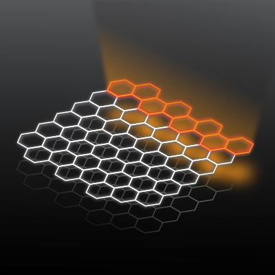
One of the main advantages of the IR spectroscopy analysis is the possibility of identifying the composition of materials by their natural vibrational response. In the case of organic biological materials, this means no need for chemical markers or fluorescent labeling for the analytical modality. Despite this asset for chemical-contrast microscopy, the long wavelengths of the mid-IR range (2.4 – 12 µm) limits IR microscopy to spatial resolutions comparable to the wavelength (Abbe limit). To overcome this limitation, scattering Scanning Near-field Optical Microscopy (s-SNOM) is an ultra-microscopy modality that allows IR spectral-imaging at the nanoscale, beyond the diffraction limit.
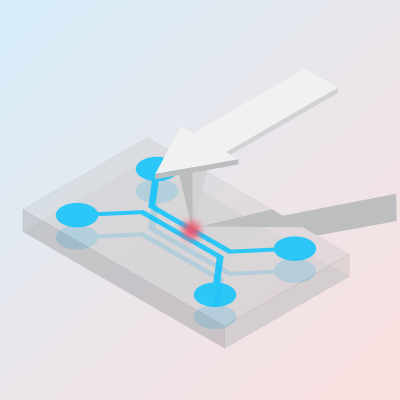
As illustrated below, the s-SNOM technique combines IR microscopy and Atomic Force Microscopy (AFM), where a metallic tip converts free-space radiation into evanescent fields at its apex, with confinement dimensions comparable to the tip radius (typically few nanometers). The technique allows accessing information on IR absorption, IR reflectivity and morphology properties of multidisciplinary materials, simultaneously.

s-SNOM technique: morphology and complex refractive index at the nanoscale
The IMBUIA-nano station combines s-SNOM and IR synchrotron in the experiment called Synchrotron Infrared Nanospectroscopy (SINS), which explores the high spatial resolution of s-SNOM (~25 nm) with the ultrawide energy coverage of the synchrotron source (mid- to far-IR). Moreover, the station is equipped with additional lasers sources allowing narrowband tunable IR imaging (QCL sources). The near-field nanoscope allows illumination/detection from both sides, therefore, it is possible to combine broadband and narrowband spectral-imaging modalities, as presented below.

Synchrotron s-SNOM combined with lasers sources. The broadband nanospectroscopy setup (synchrotron and broadband laser) enables IR spectroscopy at the nanoscale while the narrowband nanoimaging side (single line laser sources) allows for fast IR absorption/scattering imaging with nanoscale pixel size. The station is equipped with a commercial near-field nanoscope. Asymmetric Michelson interferometry (BB stage) and Pseudo-heterodyne detection (PH stage) provide for broadband interferograms and single-line complex optical response of the materials, respectively.
Broadband sources, such as synchrotrons and lasers, enable full spectral analysis in “single-shot like” experiments based on Fourier Transform (FT) interferometry and data processing. Throughput, multiplex and enhanced signal-to-noise ratio (SNR) are established advantages of FTIR, which can be explored at the nanoscale when applied to s-SNOM. The IMBUIA-nano operates with synchrotron IR from the Sirius accelerator which covers the whole mid-IR range continuously. In the broadband nanospectroscopy mode, the station enables the analysis of specific locations (point-spectrum), IR response across interfaces (spectral-linescan) and area scans (hyperspectral mapping), as organized below.

Broadband spectral coverage and analytical spectral-nanoimaging modalities available at the IMBUIA-nano station.
For fast imaging at specific frequencies, the station offers a Quantum Cascade Laser (QCL) equipped with 4 chips that are able to partially cover the IR fingerprint range. Single-line diode and gas (HeNe) lasers are also available for nano-imaging experiments within the visible spectrum.

Tunable narrowband spectral coverage and single-line nanoimaging available at the IMBUIA-nano station.
Sample preparation is an essential step for a successful analysis. Therefore, we recommend contacting the beamline staff for more information on how to prepare samples for the different modalities available. Below we present a brief guide with typical samples requirements and respective analysis methods.

Typical samples requirements for the different analytical modalities at IMBUIA.

Antibiotic resistant pathogens are a modern threat to the human health. As a worldwide spreading problem, there is an urgency for new strategies to minimize antibiotic resistance, particularly the super-resistant strains. In this work, the efficient design of carbohydrate-coated silica nanoparticles was reported which specifically targeted Gram-negative bacteria cells. Local interaction between nanoparticles and the bacterium membrane was experimentally accessed by SINS at the biomolecular level unveiling a short-range chemical connection. Thus, this novel outer membrane-targeting platform provides a new strategy to reduce drug intake and, hence, minimizes bacterial resistance.
Publication:

Lead-based organic-inorganic hybrid perovskite (OIHP) solar cells can attain efficiencies over 20%. However, the impact of ion mobility and/or organic depletion, structural changes, and segregation under operating conditions urge for decisive and more accurate investigations. Hence, the development of analytical tools for accessing the grain-to-grain OIHP chemistry is of great relevance. Here, synchrotron infrared nanospectroscopy (nano-FTIR) was used to map individual nanograins in OIHP films. The results reveal a spatial heterogeneity of the vibrational activity associated to the nanoscale chemical diversity of isolated grains. It was possible to map the chemistry of individual grains in CsFAMA and FAMA films, with information on their local composition. The analysis herein can be extended to any OIHP films where organic cation depletion/accumulation can be used as a chemical label to study composition.
Publication:
Light−matter interaction in two-dimensional photonic or phononic materials allows for the confinement and manipulation of free-space radiation at sub-wavelength scales. Most notably, the van der Waals heterostructure composed of graphene (G) and hexagonal boron nitride (hBN) provides for gate-tunable hybrid hyperbolic plasmon phonon-polaritons (HP3). Here, we presented the anisotropic flow control and gate-voltage modulation of HP3 modes in G-hBN on an air−Au micro structured substrate. Using SINS, we launched HP3 waves in hBN and observed directional propagation across in-plane heterointerfaces of the device. Our findings augment the degree of control of polaritons in G-hBN and related hyperbolic metamaterial nanostructures, bringing new opportunities for on-chip nano-optics communication and computing.
Publication:
Other theme-related publications:
A recurring goal in biology and biomedicine research is to access the biochemistry of biological processes in liquids that represent the environmental conditions of living organisms. These demands are becoming even more specific as microscopy techniques are fast evolving to the era of single cells analysis. In phase with those challenges, on-chip liquid cells emerge as a versatile alternative to control the water thickness while providing a biocompatible chemical environment for analytical analyzes. In this work we reported the development of a liquid platform specially designed for nanoscale infrared analysis of biomaterials in wet environments. A key advantage of our designed platform is the use of graphene as the optical window that interfaces wet and dry environments in the liquid cell. With SINS, we measured nanoscale fingerprint IR absorbance of a variety of liquids often used in biological studies. Further, we demonstrate the feasibility of the platform for the chemical analysis of protein clusters immersed in water with a clear view of the proteins secondary structure signatures. The simplicity of the proposed platform combined to the high quality of our data make our findings a template for future microfluidic devices targeting dynamical nanoscale-resolved chemical analysis.
Publication:

Hyperbolic phonon polaritons have recently attracted considerable attention in nanophotonics mostly due to their intrinsic strong electromagnetic field confinement, ultraslow polariton group velocities, and long lifetimes. Here we introduced tin oxide (SnO2) nanobelts as a photonic platform for the transport of surface and volume phonon polaritons in the mid to far-infrared frequency range. The report brings a comprehensive description of the polaritonic properties of SnO2 as a nanometer-sized dielectric and also as an engineered material in the form of a waveguide. By combining accelerator-based IR-THz sources (synchrotron and free-electron laser) with s-SNOM, we employed nanoscale far-infrared hyperspectral-imaging to uncover a Fabry–Perot cavity mechanism in SnO2 nanobelts via direct detection of phonon-polariton standing waves. Our experimental findings are accurately supported by notable convergence between theory and numerical simulations. Thus, the SnO2 is confirmed as a natural hyperbolic material with unique photonic properties essential for future applications involving subdiffractional light traffic and detection in the far infrared range.
Publication:

In the xylem of angiosperm plants, microscopic pits through the secondary cell walls connect the water-conducting vessels. Cellulosic meshes originated from primary walls and middle lamella between adjacent vessels, called pit membrane, separates one conduit from another. The intricate structure of the nano-sized pores in pit membranes enables the passage of water under negative pressure without hydraulic failure due to obstruction by gas bubbles (i.e., embolism) under normal conditions or mild drought stress. Here, we characterized the chemical composition of cell wall structures by SINS and AFM-IR with high spatial resolution. Characteristic peaks of cellulose, phenolic compounds, and proteins were found in the intervessel pit membrane of P. nigra wood. The advances presented here pave the way for further label-free studies related to the nano-chemistry of plant cell components.
Publication:

Infrared vibrational scattering scanning nearfield optical microscopy (s-SNOM) has emerged as a new frontier in imaging science due to its potential to provide nanoscale spatially resolved chemical spectroscopy for the investigation of molecular, soft-matter, and biological materials. As a phase-sensitive technique able to yield the full complex dielectric function of materials, different interferometric schemes have been developed involving asymmetric interferometry between sample and reference arms. In this work, we take advantage of a greatly simplified symmetric geometry that uses the spatially coherent background scattered light from within the confocal sample volume as a reference field for signal amplification in both self-homodyne and self-heterodyne interferometry. On the basis of a simple model for tip−sample scattering and interferometric detection, we demonstrate the measurement of the vibrational response of molecular materials in good agreement with established values. In addition to a compact design, enhanced signal levels, and a reduced sensitivity to fluctuations and drift, including those from the light source, self-referenced interferometry brings benefits for routine s-SNOM chemical spectroscopy, remaining robust even under a wide range of challenging experimental environments.
Publication: