CONTACT & STAFF
For more information on this beamline, contact us.


The PGM (Planar Grating Monochromator) beamline is an experimental station dedicated to X-ray Spectroscopy in the soft X-rays (100 to 1500 eV) energy range, with applications to the study of the electronic, magnetic and structural properties of materials. It is well equipped with in-situ preparation facilities, making it particularly suited for surface science and thin films characterization. In addition to that, it is offers instrumentation for microscopy and photoemission on liquids.
PGM’s source is an elliptical polarization undulator with a 50 mm period (EPU50), which allows the photon polarization to be switched among linear horizontal, linear vertical or circular polarization. Its optics is based on a standard planar grating monochromator (PGM) equipped with a 1500 l/mm variable line spacing (VLS) diffraction grating. It serves two branches that operate independently but not simultaneously.
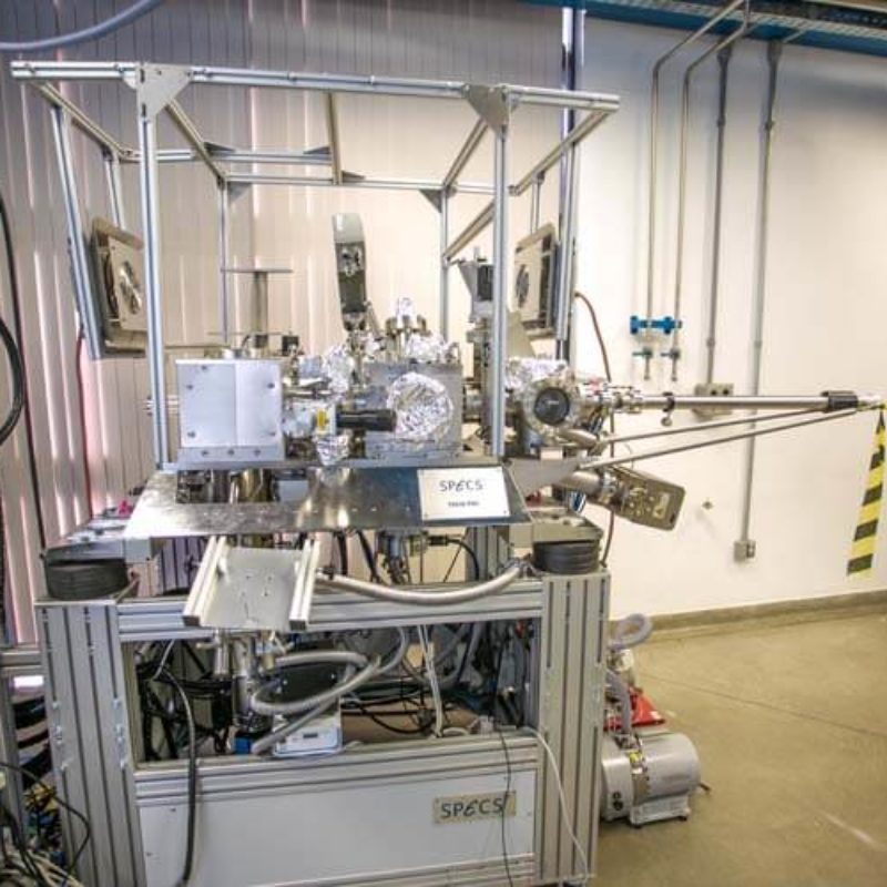
The beamline provides several experimental end stations to users. Systems are available for X-ray absorption and its variants (linear and circular dichroism), photoemission (XPS and ARPES), photoemission microscopy (PEEM), XPS on liquids and soft X-ray diffraction.
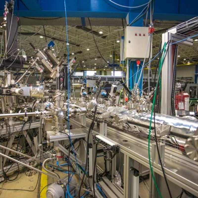
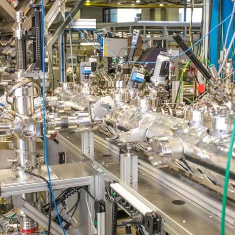
Furthermore, the beam line is equipped with a full featured in-situ preparation system, which allows the user to grow thin films and heterostructures by molecular beam epitaxy (MBE) with thermal evaporators and by pulsed laser deposition (PLD). The samples can then be transferred under ultra-high vacuum conditions and be characterized by the techniques available at the beam line.
For more information on this beamline, contact us.
The following experimental techniques and setups are available to users in this beamline. To learn more about the techniques’ limitations and requirements (sample, environment, etc.) contact the beamline coordinator before submitting your proposal.
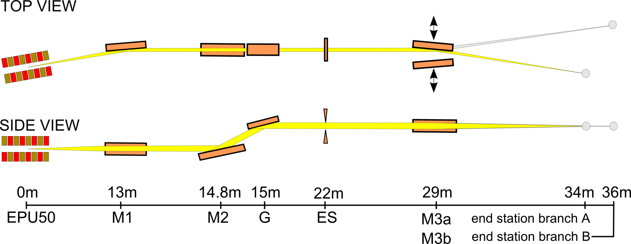
| Element | Type | Position [m] | Description |
|---|---|---|---|
| Source | Insertion Device | Insertion Device U11A, APPLE II Elliptical Polarization Undulator (EPU50), built in-house, 50 mm period, 2.73 m long | |
| M1 | Toroidal mirror | 13 | Au coated; water cooled |
| M2-G | Monochromator | 15 | Au coated plane mirror and 1500 l/mm VLS grating |
| ES | Exit slit | 22 | – |
| M3a, M3b | Refocussing toroidal mirrors | 29 | Au coated; used to switch between branches |
| Parameter | Value | Condition |
|---|---|---|
| Energy range [keV] | 103 – 1500 eV | Linear horizontal polarization |
| Energy range [keV] | 192 – 1500 eV | Linear vertical polarization |
| Energy range [keV] | 127 – 1500 eV | Circular polarization |
| Resolving power [E/ΔE] | 1000 – 25000 | – |
| Beam size at sample [mm2, FWHM] | 0.1 x 0.5 | Vertical x horizontal |
| Flux at sample [ph/s] | 1011– 1013 | – |
| Instrument | Type | Model | Manufacturer | Specifications |
|---|---|---|---|---|
| Superconducting magnet | – | – | ± 4T along the beam direction. Sample temperature between 10 and 420 K | Cryomagnetics Inc. |
| Electron Analyzer | – | – | 150 mm radius hemispherical analyzer; CCD detector Energy resolution ~2.5 meV; Angular resolution ~0.1 degree; Sample manipulator with six degrees of freedom; Sample temperature between 15 and 420 K. | Phoibos 150 SPECS |
| Photoelectron Microscopy | – | – | Fields of view from 100 down to 0.7 µm; Sample temperature from 90 up to 1400 K during measurements; Some possibilities for in-situ preparation (Ar sputtering and e-beam evaporation). | P 90 SPECS |
| Electron Analyzer for liquids sample | – | – | – | Scienta |
| Diffractometer | – | – | 4 degree of freedom diffractometer | LNLS in-house development |
All beamline controls are done through EPICS (Experimental Physics and Industrial Control System), running on a PXI from National Instruments. The data acquisition is done using a Red Hat workstation with the Py4Syn, developed at LNLS by SOL group. CSS (Control System Studio) is used as a graphical interface to display and control the beamline devices. The systems from SPECS GmbH run on the manufacturer software.
Users are required to acknowledge the use of LNLS facilities in any paper, conference presentation, thesis and any other published material that uses data obtained in the execution of their proposal.
Scientific publications produced with data obtained at the facilities of this beamline, and published in journals indexed by the Web of Science, are listed below.
Faria, M. V. G. ;Soares, E. A.;Antoniazzi, I. ;Paniago, R.M.;Miwa, R. H.;Lopes, J. M. J.;Malachias, A.;Oliveira Jr., M. H.. Experimental evidence of a mixed amorphous-crystalline graphene/SiC interface due to oxygen-intercalation, Surfaces and Interfaces, v.30, p. 101906, 2022. DOI:10.1016/j.surfin.2022.101906
Brandão, J. ;Dugato, D. A. ;Santos, M. V. P. dos;Béron, F.;Cezar, J. C.. Tuning isolated zero-field skyrmions and spin spirals at room-temperature in synthetic ferrimagnetic multilayers, Applied Surface Science, v. 585, p.152598, 2022. DOI:10.1016/j.apsusc.2022.152598
Savi, E. de L. ;Muniz, R. F. ;Silva, Jr., A. A. da ;Schiavon, G. J. ;Berrar, J. W. ;Estrada, F. R.;Schio, P.;Cezar, J. C.;Rohling, J. H.;Zanuto, V. S. ;Bento, A. C.;Medina Neto, A.;Nunes, L. A. de O.;Baesso, M. L.. Thin-film of Nd3+-Yb3+ co-doped low silica calcium aluminosilicate glass grown by a laser deposition technique, Journal of Applied Physics, v.131, n.5, p. 055304, 2022. DOI:10.1063/5.0067794
Annese, E.;Alí, A.;Barreto, J.;Felix, G. ;Stavale Jr. , F. L.. Unraveling hausmannite (Mn3O4) thin films surface structure by X ray linear dichroism, Applied Surface Science, v.578, p.151944, 2022. DOI:10.1016/j.apsusc.2021.151944
Hurtarte, I. C. C.; Amorim, H. C. S. ; Kruse, J.; Cezar, J. C.; Klysubun, W. ; Prietzel, J.. A Novel Approach for the Quantification of Different Inorganic and Organic Phosphorus Compounds in Environmental Samples by P L-2,L-3-Edge X-ray Absorption Near-Edge Structure (XANES) Spectroscopy, Environmental Science & Technology, v. 54, n, 5, p. 2812-2820, 2020. DOI:10.1021/acs.est.9b07018
Annese, E.; Mori, T. J. A.; Schio, P.; Rache Salles, B.; Cezar, J. C.. Fe-Phthalocyanine Nanoclusters on La0.67Sr0.33MnO3 Ferromagnetic Substrate for Spintronics Application, ACS Applied Nano Materials, v. 3, n. 2, p. 1516-1525, 2020. DOI:10.1021/acsanm.9b02319
Yelpo, C. ; Favre, S. ; Ariosa, D.. Detection by XRD of hidden defects in epitaxial Bi2Sr2CaCu2O8 thin films grown by PLD, Materials Chemistry and Physics, v. 239, p. 122020, 2020. DOI:10.1016/j.matchemphys.2019.122020
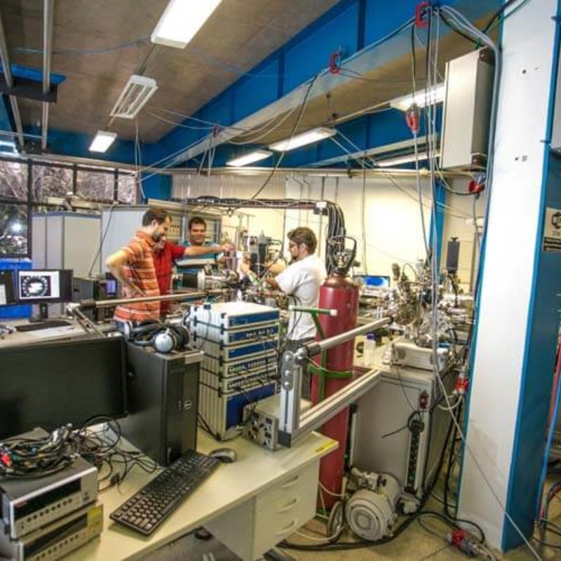
Português:
Visão geral das estações experimentais da Linha de Luz PGM.
English:
Overview of the experimental stations of the PGM beamline.
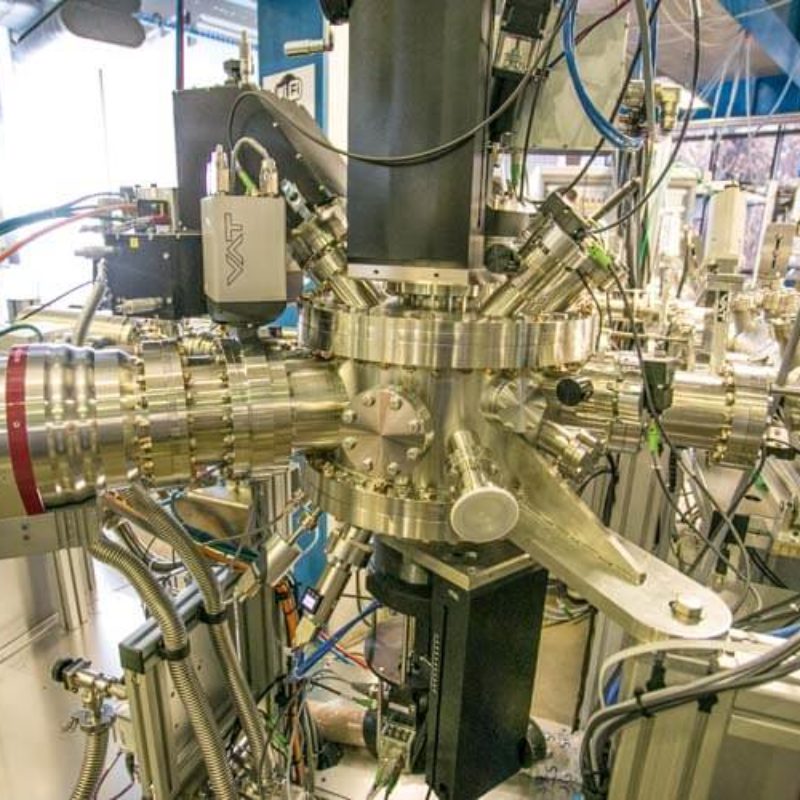
Português:
Túnel de transferência sob ultra alto vácuo.
English:
Transference Tunnel under ultra-high vacuum.
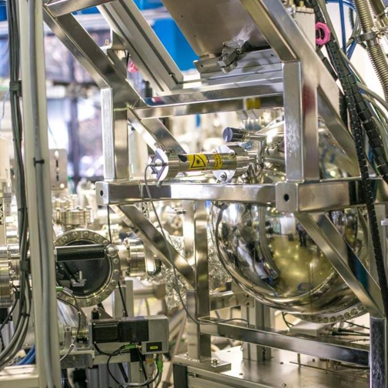
Português:
Microscópio de Fotoemissão (PEEM).
English:
Photo-electron Emission Microscope (PEEM).
Português:
Visão geral das estações experimentais da Linha de Luz PGM.
English:
Overview of the experimental stations of the PGM beamline.
Português:
Estação de fotoemissão com resolução angular (ARPES).
English:
Angle-resolved photoemission spectroscopy (ARPES) station.
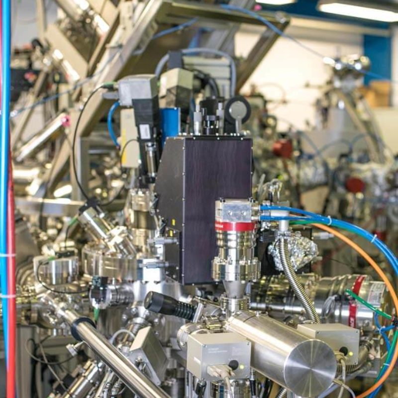
Português:
Sistema de deposição de filmes por laser pulsado (PLD, Pulsed Laser Deposition).
English:
Pulsed Laser Deposition (PLD) system.
Português:
Sistema de deposição de filmes por laser pulsado (PLD, Pulsed Laser Deposition)
English:
Pulsed Laser Deposition (PLD) system.