CONTACT & STAFF
For more information on this beamline, contact us.


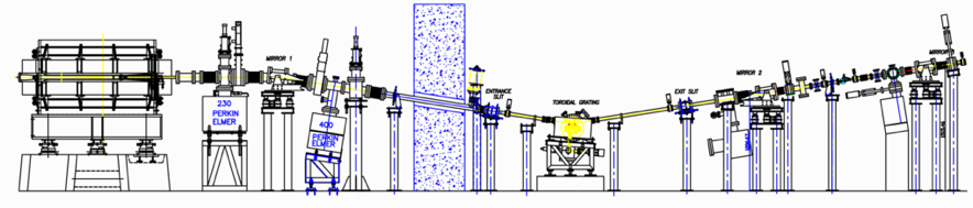
The TGM (Toroidal Grating Monochromator) beamline is dedicated to ultraviolet spectroscopy techniques in the energy range of 3 to 330 eV (ca. 400 to 4 nm). At this energy range, it is possible to perform studies related to the electronic structure and luminescence properties of solids, as well as on gas and solids subjected to conditions mimicking the atmospheric and astrophysical environments.
The TGM beamline was the first beamline to be built and made available for the users` community at the UVX storage ring of LNLS, and it has been in continuous operation since 1997. It is a spectroscopy beamline, based on a 1.67T bending magnet. It has a monochromator with three toroidal gratings and currently operates from 3 eV (413.28 nm) to 330 eV (3.75 nm), in ultra-high vacuum conditions. To ensure energy purity on the spectrum, it counts with a differentially-pumped gas filter (He, Ne, Ar, Kr), with different upper-energy cutoffs on the ionization threshold of the gases (24.6, 21.6, 15.7 and 14.1 eV, respectively), in addition to solid-state filters (glass, quartz and MgF2) for lowers energies (4.1, 8.2, 10.9 eV, respectively). Above 50 eV, the geometry of the beamline acts as an efficient cutoff of higher harmonics.
This beamline covers an important range of energy for studies of X-ray absorption of very light elements such as the K-edge of Li and the L-edge of environmentally significant ones (P, S, Cl, K). It is possible to directly assess the electronic structure of semi-conductors and insulators, to measure the luminescence of solids, photodegradation and photoionization of polymers and biomolecules, simulation of space and astrophysical conditions, and to do mass spectrometry.
Future planned experimental stations for this beamline include a Photoemission Microscope (PEEM), Circular Dichroism on the UV, for structural biology, and a dedicated chamber for optical measurements, including a setup for cryogenic studies.
For more information on this beamline, contact us.
The following experimental techniques and setups are available to users in this beamline. To learn more about the techniques’ limitations and requirements (sample, environment, etc.) contact the beamline coordinator before submitting your proposal.
Photoluminescence (PL) in the vacuum ultraviolet energy range is used to investigate the region of the optical band gap and valence band in crystalline solids. Using the PL technique, it is possible to describe the valence band, the band gap length, the position of energy levels and understanding their role during the optical process. This information may allow the development of new and customized materials for specific applications, such as biomarkers, radiation detectors, lasers, light emitting diodes (LED), phosphors for illumination devices, energy-harvesting materials, etc.
Setup: The PL setup is composed by two main modes of detection. For the first one, the excitation mode, an optical fiber is positioned in angle with the sample and the fiber cable is coupled in a photomultiplier (PMT) for integrating the light emitted by the sample after the interaction with the synchrotron radiation. This signal gives information of the excitation of the sample in a specific range of energy. The second setup is the emission one. It is similar to the first and the difference is that the optical fiber is now coupled to a spectrometer (200 – 900 nm), allowing the retrieval of the profile of the emission spectra after excitation at specific energies. The flexible setup of the detection/control of the beamline allows different complementary setups: (i) monitor the emission intensity as a function of both emission and excitation energies; (ii) time-resolved studies (for persistent and fluorescent samples). Total Electron Yield (TEY) has also been used in these studies to monitor the behavior of the sample in absorption.
Recent publications using this setup:
The beamline can be used as a light source with unique characteristics. This mode is specially interesting to cover the wavelengths below 280 nm (UVB), deeper on the UV (down to 4nm), which is present, for instance, on space conditions. In addition to producing monochromatic light, the beamline can operate in white-beam mode (full spectrum) and pink-beam mode (white-beam with the low-pass gas filter, to introduce a cutoff on the higher energies). These modes can be used to simulate the Solar radiation in space to test materials for the aerospace industry (specially polymers), and also as sources of light for astrophysics, astrochemistry in gas and solid phases, and to probe the resistance of biomolecules and microorganisms under space or planetary simulations, for astrobiology.
Setup: in this mode, the beamline normally operates with a standard chamber for the irradiation of the material. If needed, different measurement systems can be mounted to monitor the sample in situ and in real time, such as mass spectrometers (QMS, ToF) or other spectroscopic techniques (UV-Vis and Raman). Customized setups can be arranged if feasible, with prior contact with the beamline staff.
Recent publications using this setup:
| Element | Type | Position [m] | Description |
|---|---|---|---|
| SOURCE | Bending Magnet | – | Bending Magnet D05 exit A (4°), 1.67T, |
| M1 | Toroidal focusing mirror | – | R = 93.23m |
| Monochromator | Toroidal gratings, grazing incidence | – | Grating 1: 3 – 13 eV (Pt, 75 l/mm) Grating 2: 13 – 100 eV (Au, 200 l/mm) Grating 3: 100 – 330 eV (Au, 1800 l/mm) |
| M2 | Toroidal focusing mirror | – | R = 139.16m |
| M3 | Toroidal focusing mirror | – | R = 74.00m |
| Harmonics filters | Gas and solids | – | Differentially pumped gas filter (up to 24.59 eV) Solid state filters: Glass, quartz and MgF2 windows |
| Parameter | Value | Condition |
|---|---|---|
| Energy range [eV] | 3 – 330 | – |
| Energy resolution [ΔE/E] | 500 -700 | effective |
| Beam size at sample [mm2, FWHM] | 1.0 | 2mm X 0.5mm |
| Flux density at sample [ph/s/mm2] | 109 | at 10 eV |
| Flux density at sample [ph/s/mm2] | 1011 | whitebeam |
| Polarization control | – | Entrance polarization slits (upper and lower) to use natural polarization. |
| Instrument | Type | Model | Manufacturer | Specifications |
|---|---|---|---|---|
| Detector | Photomultipliers | R928 R316 R594 | – | Hamamatsu |
| Detector | Photodiodes | AXUV/SXUV100 | – | International Radiation Detectors (IRD) |
| Detector | Silicon Photomultiplier (Si-PM) | S13360-3025CS | – | Hamamatsu |
| Detector | Emission spectrograph | QE65000 | – | Ocean optics |
| Detector | Total electron Yield (TEY) | 6514 and 6485 | – | Keithley |
| Detector | VUV ionization chamber | – | – | – |
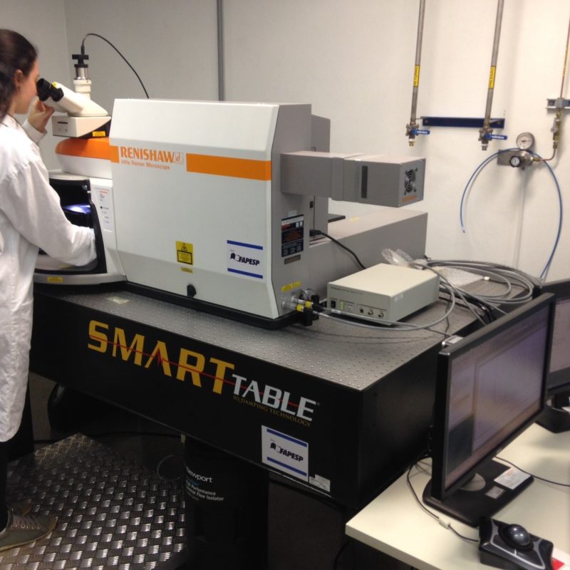
| Spectrometer | Micro-Raman for off-line measurements | inVia | Excitation lasers: 532, 633 and 785nm; Detector: CCD; Fast 2D and 3D mapping system; Fiber optics proves for in-process measurements; 5, 20, 50 and 100X objectives; Linkam temperature cell (-196°C – 600°C) | Renishaw |
The beamline is controlled by EPICS, under Linux based systems, with most of the scripts in Python and a user-friendly graphic interface.
Click here to download the TGM beamline manual (in portuguese).
Users are required to acknowledge the use of LNLS facilities in any paper, conference presentation, thesis and any other published material that uses data obtained in the execution of their proposal.
Scientific publications produced with data obtained at the facilities of this beamline, and published in journals indexed by the Web of Science, are listed below.
Hilario, E. G. ;Rodrigues, L. C. V.;Caiut, J.M.A.. Spectroscopic study of the 4f (n-1)5d transitions of LaPO4 doped with Pr3+ or co-doped with Pr3+ and Gd3+ in the vacuum ultra violet region, Nanotechnology, v.33, n.30, p.305703, 2022. DOI:10.1088/1361-6528/ac6679
Oliveira, R. de;Guallichico, L. A. G. ;Policarpo, E.;Cadore, A. R.;Freitas, R. O.;Silva, F. M. C. da ;Teixeira, V. C.;Magalhães-Paniago, R.;Chacham, H.;Matos, M. J. de S.;Malachias, A.;Krambrock, K.;Barcelos, I. D.. High throughput investigation of an emergent and naturally abundant 2D material: Clinochlore, Applied Surface Science, v.599, p. 153959, 2022. DOI:10.1016/j.apsusc.2022.153959
Coura, R, L. C. ; Andrade, A. B.; Monteiro, T. de J.; Novais, S. M. V.; Macedo, Z. S.; Valerio, M. E. G.. Photoluminescent properties of BaF2 scintillator-polystyrene composite films under vacuum ultraviolet radiation, Materials Research Bulletin, v.135, p. 111159, 2021. DOI:10.1016/j.materresbull.2020.111159
Lago, A. F.; Rogério, D. O. de ; Farias, D. B. ; Cavasso-Filho, R. L.; Dávalos, J. Z.. Investigation of the molecular structure and VUV-induced ion dissociation dynamics of 2-azetidinone (C3H5NO), Rapid Communications in Mass Spectrometry, v.35, n.3, p. e8988, 2021. DOI:10.1002/rcm.8988
Silva, A. M. B. da ; Silveira, W. S.; Matos, T. S.; Junot, D. O.; Rezende, M. V. dos S.; Souza, D. N.. Effect of terbium and silver co-doping on the enhancement of photoluminescence in CaSO4 phosphors, Optical Materials, v.111, p. 110717, 2021. DOI:10.1016/j.optmat.2020.110717
Fitaroni, L. B.; Cacuro, T. A.; Costa, C. A. R.; Lanzoni, E. M.; Galante, D.; Araujo, J. R. de; Homem, M. G. P.; Waldman, W. R.; Cruz, S. A.. Polymeric nanowrinkles: surface modification of polypropylene films in the VUV energy range, Journal of Materials Science, v.56, n.15, p.9532-9543, 2021. DOI:10.1007/s10853-021-05879-1
Artiushenko, O. ; Zaitsev, V.; Rojano, W. J. S.; Freitas, G. A.; Nazarkovsky, M. ; Saint'Pierre, T. D. ; Kai, J.. Rationally designed dipicolinate-functionalized silica for highly efficient recovery of rare-earth elements from e-waste, Journal of Hazardous Materials, v.408, p. 124976, 2021. DOI:10.1016/j.jhazmat.2020.124976
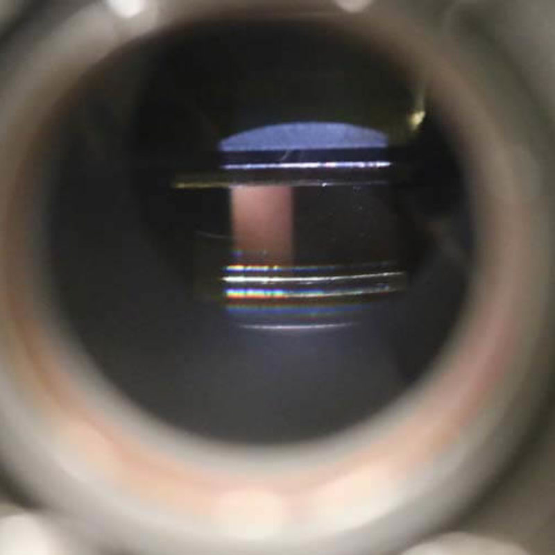
Português:
Câmara Padrão.
English:
Standard Chamber
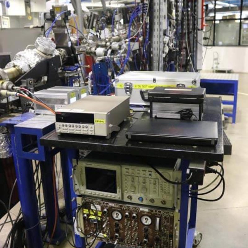
Português:
Visão geral da linha de luz TGM.
English:
Overview of the TGM beamline.
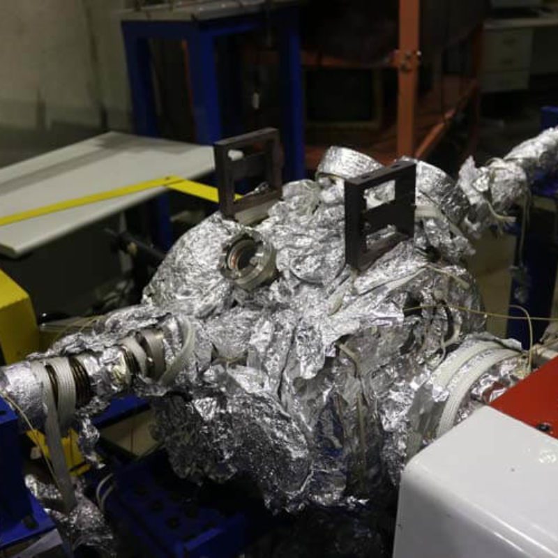
Português:
Vista lateral da Linha de Luz TGM.
English:
Lateral view of the TGM beamline.
Português:
Monocromador.
English:
Monochromator.
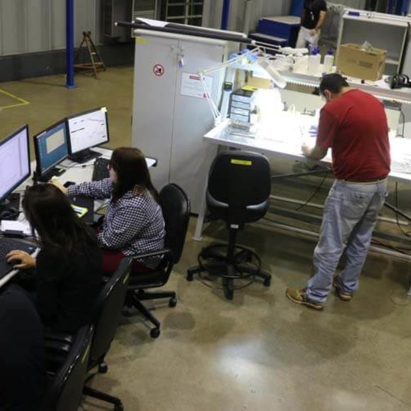
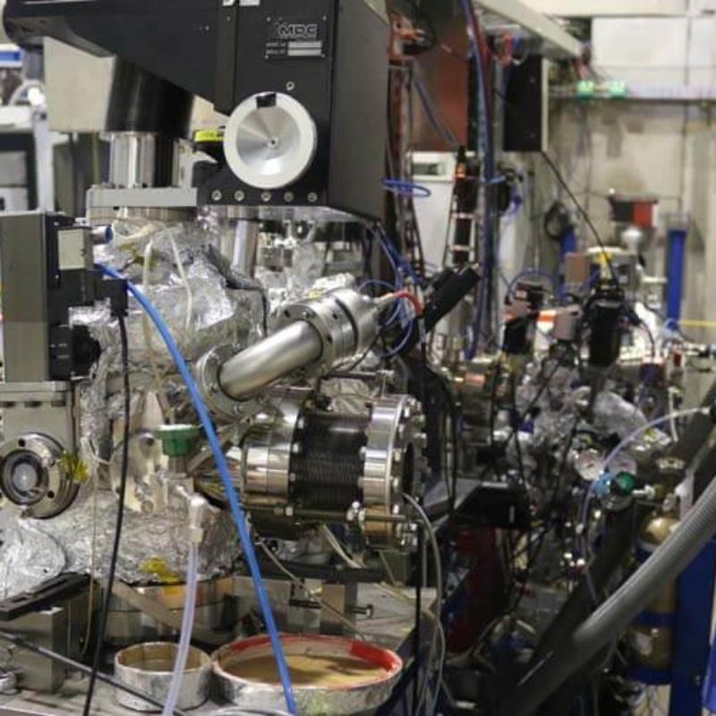
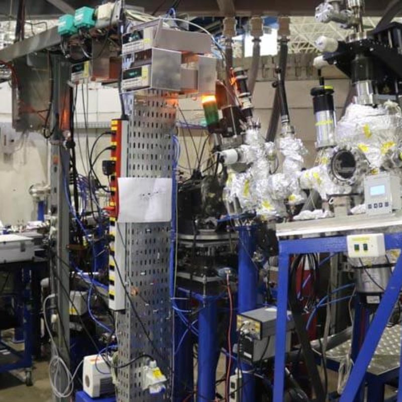
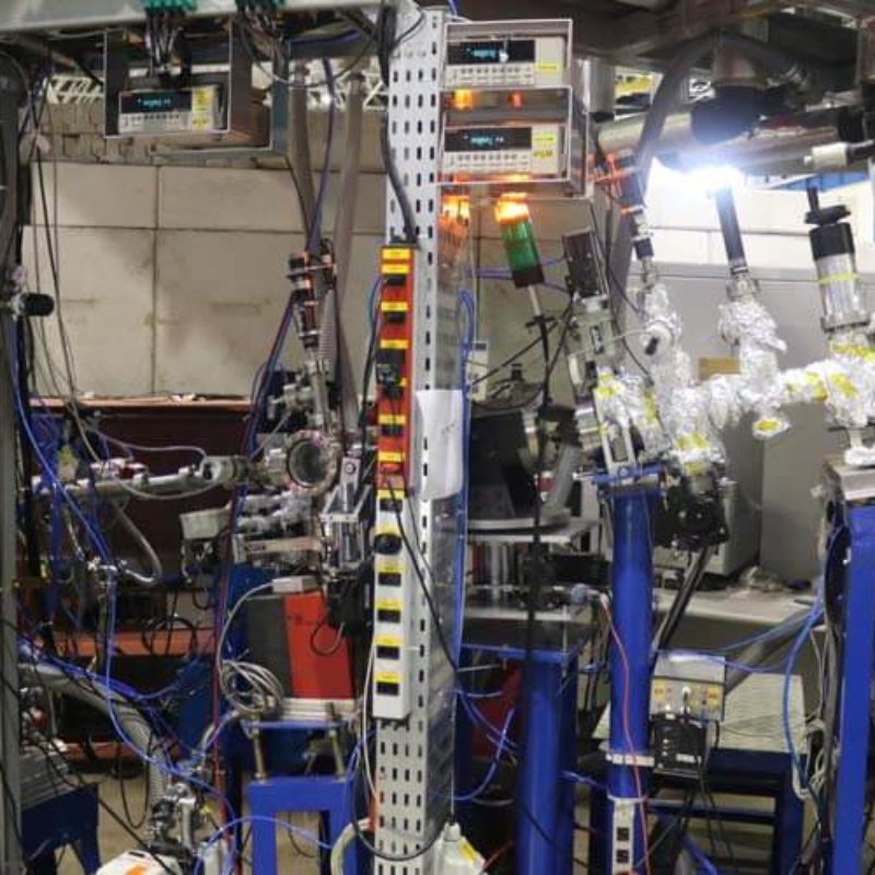
Português:
Estações de trabalho para Usuários.
English:
Workstation for Users.
Português:
Aparato para medidas ópticas.
English:
Optical measurement apparatus.
Português:
Grade de Difração no Monocromador.
English:
Diffraction gratings in the Monocromator.
Português:
Espectrômetro micro-Raman Renishaw in-Via para medidas fora da linha.
English:
Renishaw inVia micro-Raman spectrometer for off-line measurements.
Português:
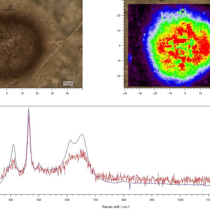
Exemplo de mapeamento Raman 2D de óxido de ferro em um microfóssil.
English:
Example of a 2D Raman mapping of iron oxide on a microfossil.
Português:
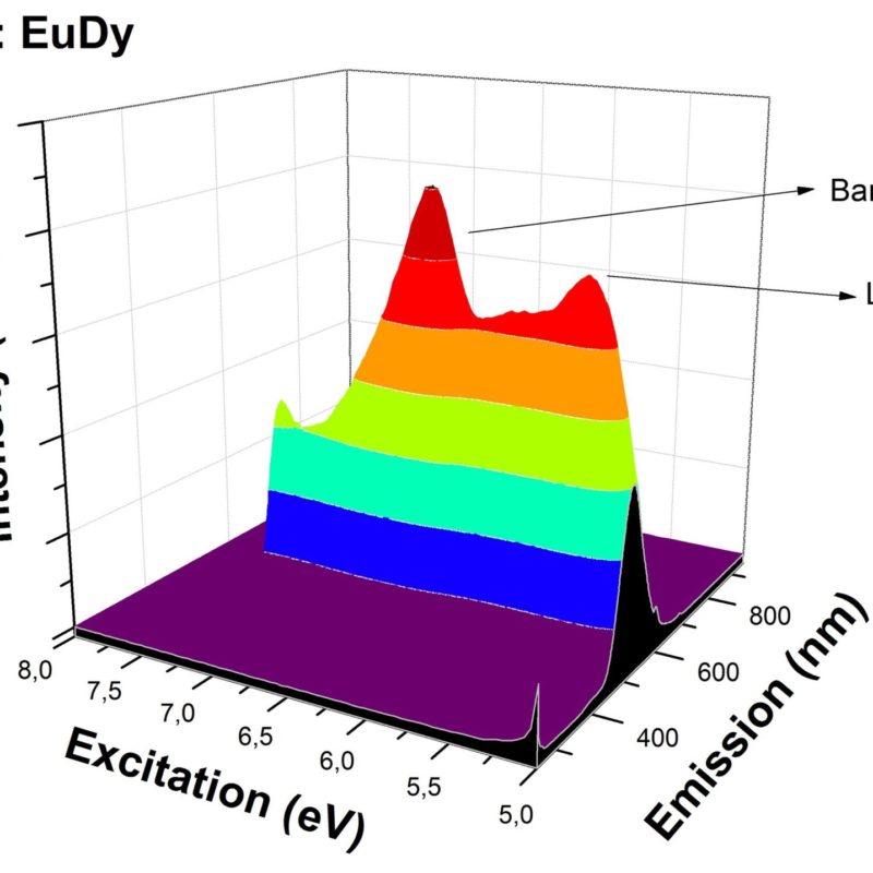
Exemplo de mapeamento 3D (energia de excitação x comprimento de onda de emissão x intensidade de emissão) da região do band gap de um aluminato de estrôncio dopado com terras raras.
English:
Example of a 3D mapping (excitation energy x emission wavelength x emission intensity) around the optical band gap of a rare earth doped strontium aluminate.
Português:
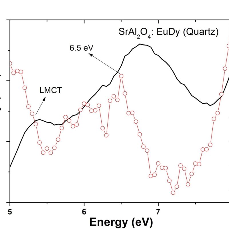
Espectro de excitação ao redor do band gap óptico de um aluminato de estrôncio dopado com terras raras.
English:
Excitation spectrum around the optical band gap of a rare earth doped strontium aluminate.
Português:
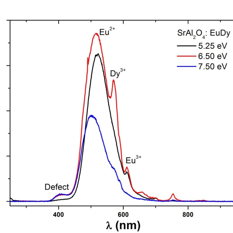
Espectros de emissão de um aluminato de estrôncio dopado com terras raras, excitados em diferentes energia no ultravioleta de vácuo.
English:
Emission spectra of a rare earth doped strontium aluminate, excited in the vacuum ultraviolet energy range.
Português:
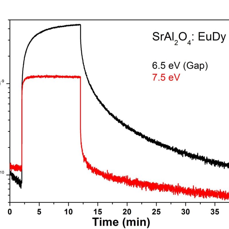
Curvas de decaimento persistente de um aluminato de estrôncio dopado com terras raras, excitadas acima e na posição do band gap do material.
English:
Persistent decay curves of a rare earth doped strontium aluminate, excited above and at the material band gap position.