

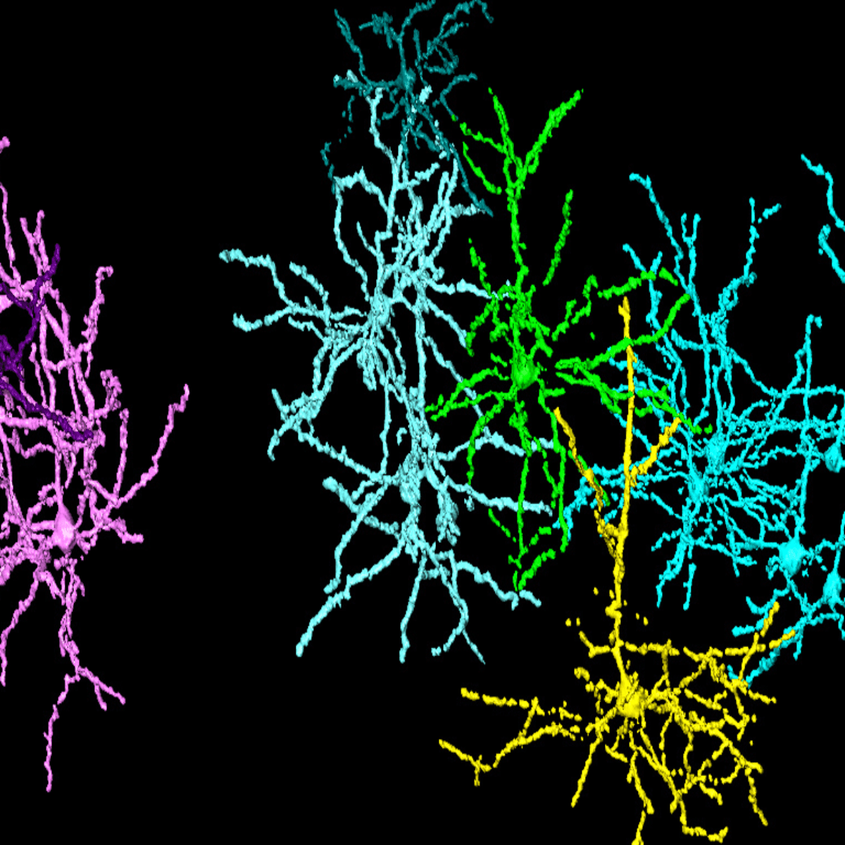
X-ray microtomography of the cerebral cortex showing the segmentation of individual neurons. Each color represents a single neuron or a group of neurons.
Results open new perspectives for the study of neurodevelopment and neurodegenerative diseases
A comprehensive understanding of the brain, its development, and eventual degeneration, depends on the assessment of neuronal number, spatial organization, and connectivity. However, the study of the brain architecture at the level of individual cells is still a major challenge in neuroscience.
In this context, Matheus de Castro Fonseca, from the Brazilian Biosciences National Laboratory (LNBio), and collaborators [1] used the facilities of the Brazilian Synchrotron Light Laboratory (LNLS) to obtain, for the first time, three-dimensional images in high resolution of part of the neuronal circuit, observed directly in the brain and with single cell resolution.
The researchers used the IMX X-Ray Microtomography beamline, in combination with the Golgi-Cox mercury-based impregnation protocol, which proved to be an efficient non-destructive tool for the study of the nervous system. The combination made it possible to observe the points of connectivity and the detailed morphology of a region of the brain, without the need for tissue slicing or clearing.
The mapping of neurons in healthy and unhealthy tissues should improve the research in neurodegenerative and neurodevelopmental diseases. As an example of this possibility, the work presents, for the first time in 3D, the neuronal death in an animal model of epilepsy.
The researchers are now working to extend the technique to animal models of Parkinson’s disease. The intention is to better understand the cellular mechanisms involved in the onset and progression of the disease. In the future, with the inauguration of the new Brazilian synchrotron light source, Sirius, the researchers believe that it will be possible to obtain images at the subcellular level, that is, images of the interior of the neurons.
Repercussion: This research was highlighted by the brazilian news media, such as the newspaper Folha de S.Paulo, and the TV news programs Jornal Nacional and Jornal da Band.
Source: [1] Matheus de Castro Fonseca, Bruno Henrique Silva Araujo, Carlos Sato Baraldi Dias, Nathaly Lopes Archilha, Dionísio Pedro Amorim Neto, Esper Cavalheiro, Harry Westfahl Jr, Antônio José Roque da Silva, Kleber Gomes Franchini, High-resolution synchrotron-based X-ray microtomography as a tool to unveil the three-dimensional neuronal architecture of the brain, Scientific Reports 8, 12074 (2018). DOI:10.1038/s41598-018-30501-x

Research investigates cheaper alternatives to attenuate the emission of toxic gases
First experimental report of a special optical layout dedicated to correct typical aberrations derived from large extraction ports in IR beamlines