

Researchers achieve unprecedented details of the shape, composition and preservation of microfossils
For decades, scientists have been using fossils of microorganisms to better understand the origin and evolution of life on Earth, but this branch of palaeobiology has taken a great leap forward with the development of novel imaging techniques. Historically, the study of the earliest traces of life on Earth has been surrounded by a lot controversy and technical challenges. Sometimes it is even difficult to tell out if a structure is really a fossil or… just an artefact.
These challenges are related to the characteristics of these microfossils: they are only few micrometers in size (ten times less than the thickness of a human hair). Also, ancient rocks have suffered some degree of geological alteration due to the pressure and temperatures exerted by layers of rock above them. Therefore, the original components of these tiny cells have been “cooked” at temperatures of more than 100°C, substituted by minerals and pressed for hundreds of millions to billions of years, before ending up in the hand of scientists.
This challenge is shared with astrobiology in the search of life on other planets, like Mars. NASA and ESA are planning a mission for returning samples from that planet in the next decade to search for signatures of life, among other objectives. A main question is: if there are fossil microorganisms on Martian rocks, how are we going to identify them? To be ready for this task, we need to develop approaches to unequivocally identify the earliest traces of life on Earth and understand the conditions that allowed them to be preserved for so long.
For this reason, researchers from the Brazilian Synchrotron Light Laboratory (LNLS), in collaboration with scientists from France and Switzerland have obtained the most detailed 3D views ever achieved of very ancient traces of life on Earth. The studied microfossils, from the Gunflint Formation, in Canada, are approximately 1.9 billion years old, and are the preserved remains of microorganisms similar to bacteria existing today, but from a period when only microscopic life existed on Earth.
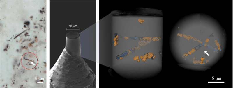
Microfossils from the Mink Mountain Locality by different imaging methods. From the left to the right: optical microscopy, scanning electron microscopy and Ptychographic X-ray computed tomography.
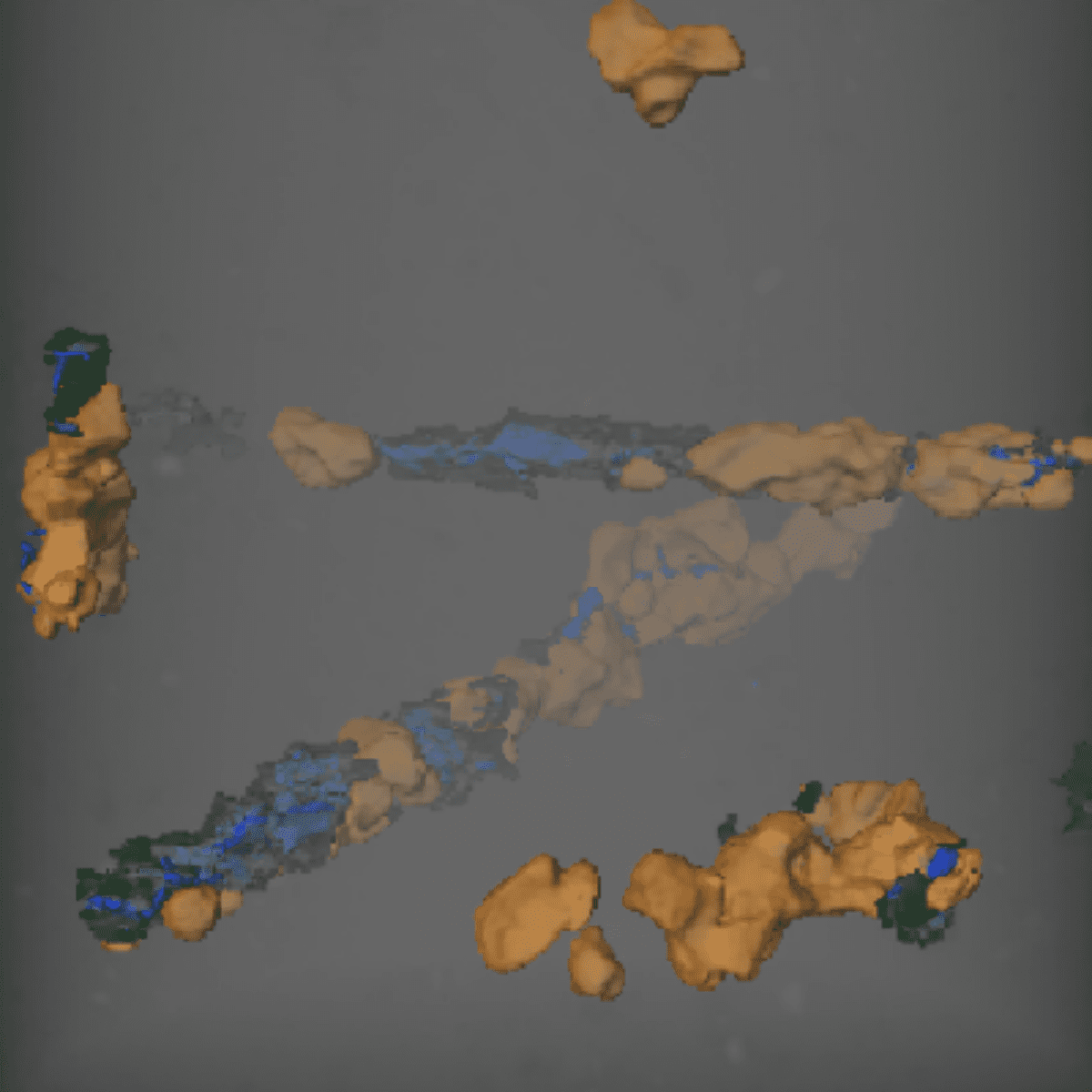
Using a state-of-the-art tomography with very high resolution called Ptychographic X-ray Computed Tomography at the Swiss Light Source, Paul Scherer Institute, Switzerland, they were able to see the microorganisms in 3D inside tiny pieces of rock without having to crack it open, and also identity materials based on their electron density. They were then able to reconstruct the cells in 3D and see how time and geological processes affected their original shape differently in two different parts of the same geological formation. In one locality, called Mink Mountain, the fossils had been previously described as hematite-coated structures, but the researchers identified masses of mature carbonaceous materials that was not observed by optical microscopy. Furthermore, instead of hematite, crystals of the less-common iron oxide maghemite were found associated with the fossils, revealing a previously unknown process involved in the preservation of these structures, and improving our comprehension of how these structures were preserved and how they were altered after being buried for billions of years.
This study also opens new and exciting possibilities for the controversies surrounding the oldest traces of life that have already been reported. Currently, synchrotron technology is taking a new step with the onset of 4th generation accelerators, such as Sirius, in Brazil, and MAX IV in Sweden, and the upgraded 3rd generation machines like the upgraded ESRF in Grenoble, and further SOLEIL, in Paris-Saclay, and the Swiss synchrotron SLS, both under preparation. These sophisticated machines have the potential to reveal further novel and exciting details about the earliest traces of life on Earth or even from Mars, and might help us answer some of the most intriguing questions of science: how did life appear on Earth? And are we alone on the Universe?
Sirius
At Sirius, the Carnaúba beamline will allow experiments to be carried out using ultra-high resolution tomography, with great potential for the study of microfossils. The technique can be applied to various types of materials, including living cells, which may have their internal structure detailed in-vivo, allowing, for example, the development of more efficient drugs. The area of materials science may also make a leap in quality, now that it will be possible to reveal the structure and composition of metallic alloys, ceramics, and many other compounds of technological interest. The technique should also enable the development of more efficient oil exploration methods by characterizing the tiny pores of the rocks where the oil is trapped.
Source: Maldanis, L., Hickman-Lewis, K., Verezhak, M. et al. Nanoscale 3D quantitative imaging of 1.88 Ga Gunflint microfossils reveals novel insights into taphonomic and biogenic characters. Sci Rep 10, 8163 (2020). DOI: 10.1038/s41598-020-65176-w

Research investigates new niobium-based materials for improving electrical energy storage

Research contributes to the design of more effective antibiotics and anticancer compounds