

First 3D imaging experiments at Sirius were performed using ptychographic nanotomography
Researchers from the CATERETÊ beamline have succeeded in the very first measurements of 2D and 3D imaging at Sirius, the new Brazilian synchrotron light source of the Brazilian Center for Research in Energy and Materials, a private non-profit organization under the supervision of the Brazilian Ministry of Science, Technology, and Innovations (MCTI).
The CATERETÊ beamline is dedicated to coherent and time-resolved scattering experiments. Experiments such as coherent X-ray diffractive imaging (CXDI) and X-ray photon correlation spectroscopy (XPCS) are at the core of the beamline’s activities, as well as time-resolved small-angle X-ray scattering (SAXS).
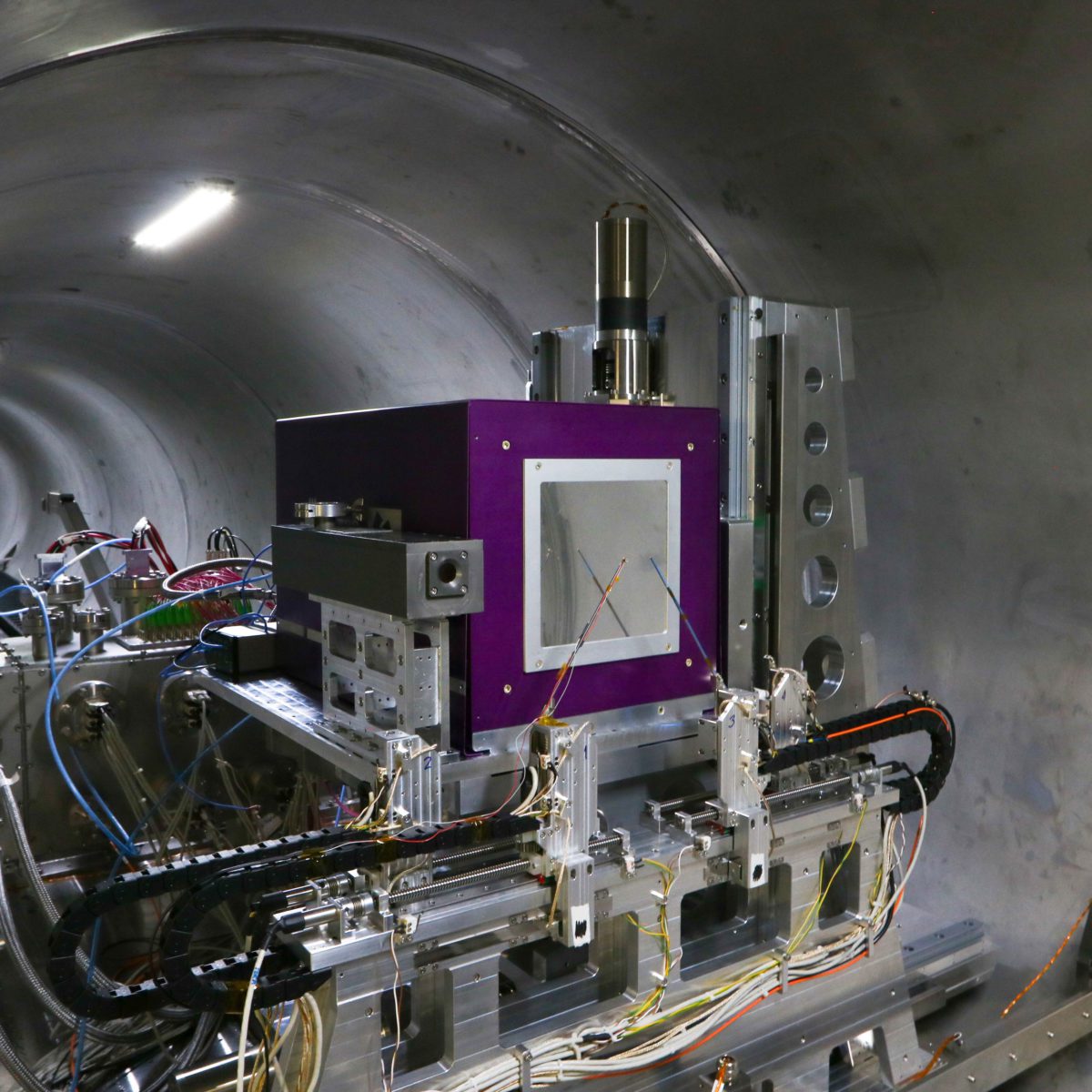
The CATERETÊ beamline is equipped with an undulator source, in a low-beta straight section, and two cryo-cooled focussing mirrors creating a 40 x 40 mm2 coherent beam at 88 m from the source. The beamline operates in the 4 to 24 keV energy range using a horizontally deflecting 4-bounce crystal monochromator (4CM). Moving the 4CM laterally by a few millimetres enables the operation of the beamline in pink beam mode, maintaining the beam position unchanged. The experimental station is located 88 m from the source, followed by a 28 m vacuum chamber (AVS, Spain) hosting the in-vacuum detector, the PIMEGA 540D (Figure 1).
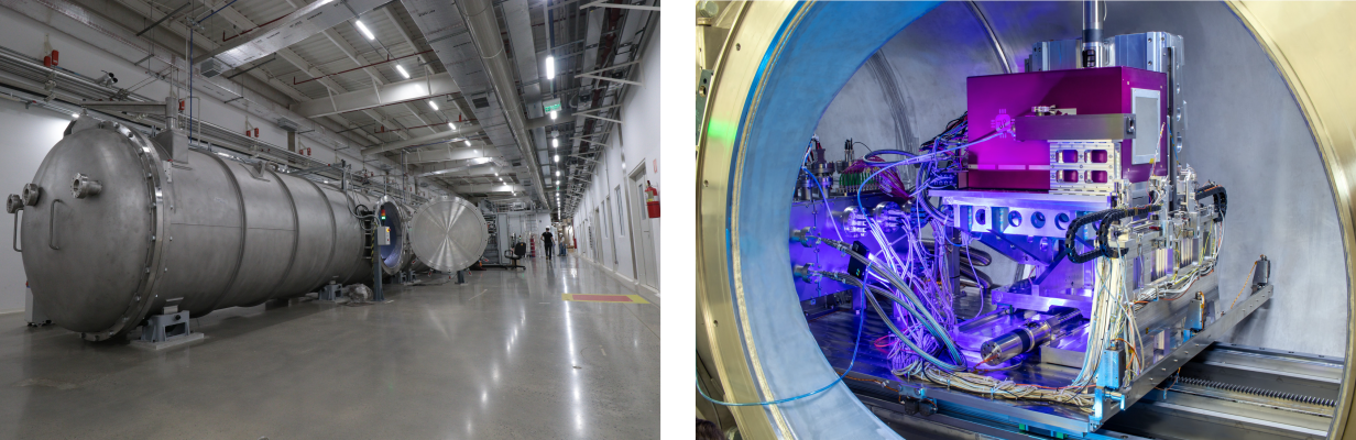
Figure 1: 28 meters vacuum tunnel (left) hosting a silicon PIMEGA 540D detector (right).
The PIMEGA family of detectors was developed in a partnership between CNPEM and Pi Tecnologia (PITEC), a company dedicated to the development of communication and imaging systems with state-of-the-art electronic systems. The detectors make use of CERN Medipix3RX ASIC, with a pixel size of 55 µm which makes direct detection of photons to deliver the highest spatial resolution data (9,4 megapixels) with a 2000 fps data frame rate.
Synchrotron X-Ray Ptychography
The measurement of Siemens Star sample using ptychography is a standard procedure when commissioning a beamline for these experiments, providing important information such as the achieved 2D resolution. Ptychography is a version of the CDI technique where a sample is raster-scanned relative to the X-ray illumination and multiple diffraction patterns are recorded in the far-field. A 3.8 keV pink beam (2% bandwidth) with 70 mA in the storage ring was used. The detector was placed 14 meters from the sample. Figure 2 shows the experimental station. The coherent beam reaches the siemens star after passing through a 5 µm pinhole. The scattered beam then travels through the vacuum tunnel (10⁻³ mBar) to the detector
The sample was raster-scanned with 1.25 µm steps to record a set of diffraction images with the PIMEGA detector at 150 ms exposures and 24-bit counter depth (Figure 2).
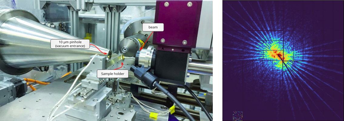
Figure 2: Left: Sample station of the experiment. Right: Scattering pattern at PIMEGA detector in a single frame.
An iterative phase-retrieval algorithm developed by LNLS’ scientific computing group was used to reconstruct the diffraction images and obtaining the 2D image of the sample under study (Figure 3).
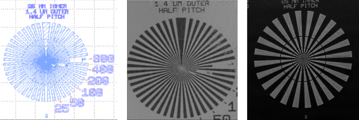
Figure 3: Left: Reference model of the sample pattern. Center: Reconstruction of the siemens star structures test pattern with multiple spoke distances in nanometers, acquired at E = 3.8 keV, sample to detector distance = 28 meters, pinhole = 10 μm, Step scans = 2 μm, field of view indicated on the reconstructions, reconstruction pixel size = 50 nm. Right: A similar experiment resulting in a smaller reconstruction pixel size. In this example, it is possible to distinguish the 25 nm features in the center of the siemens star.
The visualization of complex morphologies and hierarchical structures requires the use of advanced 3D imaging methods. In this first experiment, the CATERETÊ team explored three-dimensional porous polymeric membranes using synchrotron ptychographic broadband X-ray nanotomography (X-ray energy 3.8 keV, sample to detector 14 m, 5 µm pinhole).
The polyethersulfone sample was raster-scanned with steps of 1.25 µm. The total field of view covered in a projection was 28 x 24 μm2 and in total 460 projections were used for a tomogram reconstruction. The resulting voxel size in the reconstructed projections is 27 nm while the spatial resolution of the ptychographic tomograms were estimated by Fourier shell correlation, and lead to 33 nm. The three-dimensional renderings are presented in Figure 4.
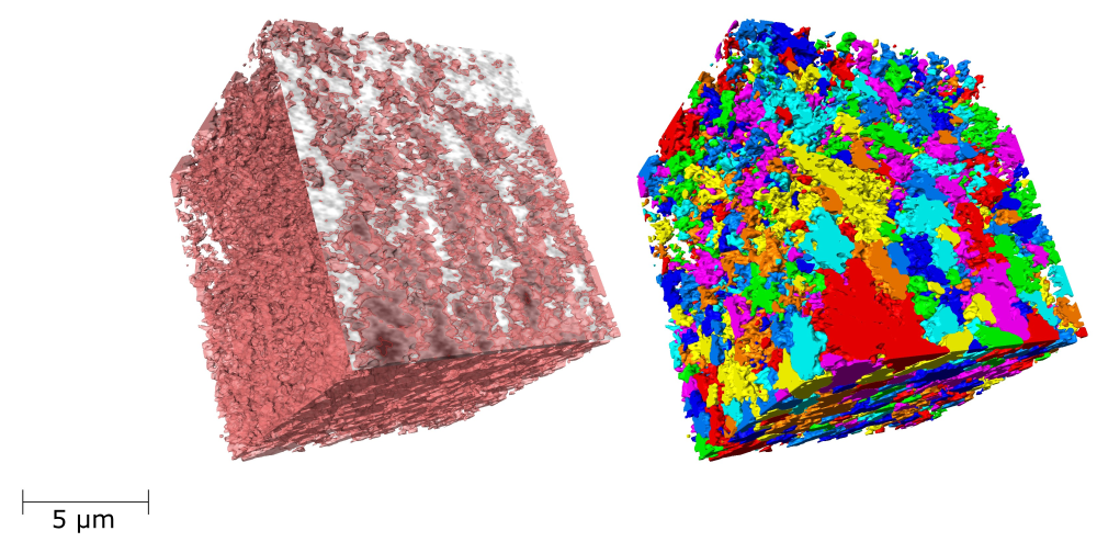
Figure 4: Ptychographic X-ray computed nanotomography of a polyethersulfone polymer membrane. Left: 3D rendering of the polymer membrane with the matrix in pink and pores in white. Right: different colors indicate the several porous networks present inside the membrane.

This call for proposals intends to supply beamtime for the users during the beamline commissioning period
LNLS is developing courses with a focus on fundamental aspects for the use of the new beamlines