

Review article was highlighted in the Applied Physics Reviews journal and explains how computed micro- and nanotomography can be used in fourth-generation synchrotrons like Sirius
Fourth-generation synchrotron light sources like Sirius expand the capacities of x-ray micro- and nanotomography to new levels. A review article written by a team of researchers from the Brazilian Center for Research in Energy and Materials (CNPEM) and the Federal University of ABC and published in Applied Physics Reviews describes recent developments in x-ray computed tomography associated with synchrotron light and artificial intelligence.
X-ray computed tomography is a medical resource that has been known in medicine since the 1970s. It is capable of producing 3D images that provide a non-invasive detailed view of various structures in the human body and offers a very valuable tool for diagnostics; it was not long before this technique was utilized in other areas of science.
The millimeter-scale resolution offered by medical tomography equipment was brought down to the micro-scale in the early 1980s and used to study biological structures, allowing quantitative morphometric analyses. The emergence of synchrotron light sources in that same decade and resulting ability to use monochromatic high-brightness X-rays continued to boost the resolution of images obtained from these techniques.
In the early 2000s, computed microtomography (μCT) began to be utilized in studies involving plants, animals, soil, and synthetic carbon-based materials, permitting notable advances in the understanding of various phenomena and systems. But the restricted capabilities of conventional x-ray sources limited the spatial resolution of the images captured, and significantly lengthened the time needed to collect data.
The emergence of fourth-generation synchrotron light sources like Sirius led to new possibilities for research in various areas. Between third- and fourth-generation light sources, brightness increased by two orders of magnitude. This, along with the high coherence of the resulting beam, makes it possible to use micro- and nanotomography image capture techniques that are much better suited to studying carbon-based materials.
The stronger stream of photons also allows these analyses to be resolved in real time: in other words, images are captured fast enough to view changes in the structure of materials over time, making in situ, in vivo, and in operando analyses feasible.
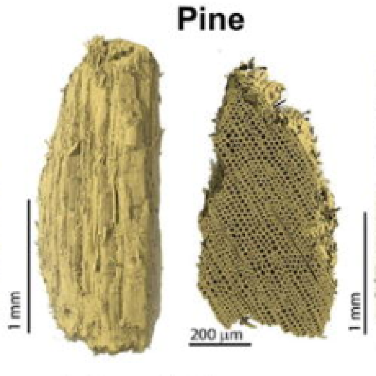
As the article by the CNPEM researchers explains, the applications of these techniques in research involving plants extend into countless fields of study. Researchers can investigate transport phenomena such as liquid permeability in various tissues, characteristics of grains and fruit that affect food quality, processes of chemical modification, adhesive application and degradation of wood, and biomass transformations to produce new chemical products and liquid biofuels.
The multidisciplinary capabilities of these analysis techniques are clear in these examples; even if we consider only research on organic matter, the applications extend from basic research in the field of biology to industrial processes for manufacturing materials and generating energy.
But x-ray tomography goes even further: since this method is non-destructive, it is ideal for field research in agriculture and the environment. Soil samples can be analyzed for classification by size, state of aggregation, porosity, and mineralogy of the solid portion, which is directly related to many different transport processes for water, nutrients, and contaminants. Similarly, the plant/soil interface can also be studied in order to understand how growth takes place and how water and nutrient absorption is regulated.
Research involving animal tissue can also put these techniques to good use. The resulting images help us to understand the processes involved in tissue development, functionality, injuries, fractures, and even disease progression.
Micro- and nanotomography of synthetic carbon-based materials (one of the main focuses of the article and also a topic of research in the area of renewable materials at the Brazilian Nanotechnology National Laboratory, LNNano), helps in the study of their 3D structures and correlation with the fabrication parameters used. And real-time analysis allows researchers to characterize these materials in situ, making it possible to observe transformations and behavior in materials when they are subjected to external stresses and fluids.
As Rubia Figueredo Gouveia, the researcher who conceived the idea for the article, notes, “these techniques allow a qualitative as well as quantitative analysis of these materials which we call morphometry. We are able to calculate parameters such as pore size, total porosity, distribution of particles in different phases, and thickness of solid walls around pores, for example.”
Despite all these benefits offered by fourth-generation synchrotrons, there are also some drawbacks. The greater temporal and spatial resolution obtained in the tomography images translates into enormous amounts of data generated, and extra difficulties in processing and quantitative analysis of these images.
While in the past the limiting factor and major challenge to researchers involved the minimal scale permitted, today it is found in the field of computation. Pedro Ivo Cunha Claro, another author of the article, emphasizes that “computers are what can limit fourth-generation synchrotrons. You need powerful computation installations to process this data. Today synchrotron light generates so much data that it is computationally challenging to process it all. We are applying artificial intelligence as a strategy to address this problem.”
Supervised and non-supervised machine learning models could help with segmenting and quantitative analysis of the high-resolution tomographic images. As the article shows, convolutional neural networks are ideal for handling large volumes of data related to computer vision. But developing these models is itself a challenge: they involve millions of parameters and require expertise in areas like artificial intelligence and computer vision, and their application may be limited to very specific domains due to problems with generalizing current techniques.
There is also one major difficulty involved in validating these models: “Determining the accuracy of results for segmenting images with nanometric resolution is a large challenge. Since we are at a previously unreached scale of resolution, the stage of generating reference images (which is generally done by specialists) can be extremely complex and in many cases is a limiting factor for us to precisely determine the accuracy of a model learned via a machine learning technique,” says Allan da Silva Pinto, a coauthor of the article and coordinator of the Data Management and Science group at the LNLS.
The computation infrastructure at Sirius already includes equipment that can help researchers manage and process this huge quantity of data, which is an important issue not only for the MOGNO beamline, which is dedicated to x-ray micro- and nanotomography, but also other beamlines. TEPUI, the throughput enhanced processing unit, serves the community of Sirius users and is an extremely important tool in the research process.
According to Nathaly Archilha, coordinator of the MOGNO beamline, Sirius already has 4 petabytes of storage; the real challenge is dealing with so much data and post-processing via quantitative analyses. “We need to create these tools. And we need internal researchers using these tools so that they can evolve rapidly and we can offer these solutions to our users.”
The Brazilian review article highlights not only the massive potential offered by fourth-generation synchrotron light sources for x-ray micro- and nanotomography, but also the need to combine multidisciplinary efforts to take full advantage of this potential.
“There are already very advanced studies on deep learning which focus on processing and image analysis, as well as others investigating x-ray micro- and nanotomography. But we don’t see these two areas in a complete dialog, which is exactly what our work addresses. This article will be a good foundation for first-time users as well as those with advanced knowledge of tomography,” adds Rubia Figueredo Gouveia.
The article published in Applied Physics Reviews involved contributions by nine researchers: Pedro Claro, Egon Borges, Gabriel Schleder, Nathaly Archilha, Allan Pinto, Murilo Carvalho, Carlos Driemeier, Adalberto Fazzio, and Rubia Gouveia, who are part of the national laboratories at the CNPEM, and was the result of FAPESP project 2020/08651-4.
Researchers at USP in São Carlos combined cutting-edge technologies and demonstrated that a molecule targeted by medications behaves differently than previously theorized.
Paper published at Science Magazine investigates unexpected molecular interactions that affect cell function and could cause disease