CONTATO & EQUIPE
Para mais informações sobre a linha de luz, entre em contato.
A linha de luz SAXS1 é dedicada ao espalhamento de raios X a baixo ângulo (SAXS), operando em uma energia fixa de 8 keV. Ela foca em investigações estruturais de materiais e amostras biológicas, na escala de nanômetros a micrômetros, com aplicações em ciência de matérias, química, géis, reologia, biologia estrutural, ciências ambientais e geociências. O SAXS permite o estudo de amostras sólidas e líquidas com ambientes em ar ou em vácuo. A detecção é feita em vácuo utilizando um detector Pilatus 300K (Dectris)
Outra técnica disponível é o espalhamento de raios X a amplos ângulos (WAXS) estudos de amostras cristalina e com as mesmas aplicações, ambientes de amostra e detecção em ar (Pilatus 100K – Dectris).
Devido ao grande fluxo de fótons, experimentos de SAXS resolvido no tempo podem ser executados, permitindo atingir resoluções de sub-segundos para estudo cinéticos.
Diversos ambientes de amostra estão disponíveis para a comunidade tais como (1) fornos (Linkam THMS600 *), permitindo atingir temperaturas de -200°C até 600°C; (2) dispositivos de estiramento (Linkam TST350); (3) bomba peristáltica para circulação de solução em experimento in situ; (4) dispositivo Stopped-flow* destinado ao estudo de amostras biológicas; (5) Autosampler (Spark Holland) para injeção automática em estudos de proteínas.
Outras técnicas experimentais disponíveis incluem o espalhamento de raios X a amplos ângulos (WAXS)
(*) Equipamentos multiusuários financiados pela FAPESP (projeto: 04/09447-9): “PROEM- Instrumentação da Linha SAXS2 do LNLS: aplicações da técnica de SAXS ao estudo de materiais nanoestruturados, polímeros densos e sistemas biológicos”.
Para mais informações sobre a linha de luz, entre em contato.
As técnicas e configurações experimentais a seguir estão disponíveis nesta linha de luz. Para saber mais sobre as limitações e requerimentos das técnicas, contate o coordenador da linha de luz antes de submeter sua proposta.
Este experimento permite a determinação de parâmetros estruturais tais como distâncias presentes nas moléculas, raio de giro, tamanhos de poros, massa, morfologia, estado de agregação etc, com dimensões que vão de 1 a > 100 nm. A tabela abaixo resume os parâmetros disponíveis na linha SAXS1.
Intervalo em q (nm-1) |
Distância amostra-detector (m) |
0,1 – 5,0 |
1,0 |
0,08 – 3,0 |
1,5 |
0,04 – 1,5 |
3,0 |
Esta técnica é especifica para amostras biológicas, permite ainda a obtenção e construção de modelos tridimensionais em baixa resolução, através da técnica ab initio. Devido a ampla faixa de intervalos em q que podem ser atingidos é possível realizar estudos de moléculas de tamanhos variados. Nestes experimentos, um Autosampler SparkHoland é usado para injeção automática de amostras biológicas.
Espalhamento de raios X a baixo ângulo resolvido no tempo (TR-SAXS)
Esta técnica permite acompanhar mudanças de tamanho e propriedade das moléculas estudadas em um dado intervalo de tempo com velocidade de aquisição de alguns milissegundos até > minutos.
Este experimento permite a determinação de estruturas de mais alta resolução de estruturas cristalinas de polímeros e fibras, uma vez que utiliza a análise dos picos de Bragg para determinação dos planos cristalinos e consequentemente distância inter-planares. Permite avaliar as modificações de propriedades estruturais pela aplicação de diferentes condições em materiais, inclusive biológico.
| Elemento | Tipo | Posição [m] | Descrição |
|---|---|---|---|
| Fonte | Dipolo de Curvatura | 0.0 | Dipolo de Curvatura D01 Saída B (15°), 1.67 T, 890 µm x 150 µm a 8 keV |
| S0 | Fendas Feixe Branco | 7.2 | |
| Mono | Monocromador Toroidal Side-bounce | 8.0 | W/B4C multi-camada (500 camadas duplas) em substrato de Si |
| S1 | Fendas | 8.5 | |
| S2 | Fendas | 14.0 | |
| S3 | Fendas | 16.0 | |
| Detector | Pilatus 300K | 17.2 |
| Parâmetro | Valor | Condição |
|---|---|---|
| Faixa de Energia [keV] | 8 | Si(111) |
| Resolução de Energia [ΔE/E] | 0.1 | Si(111) |
| Tamanho do feixe na amostra [mm2, FWHM] | 1.5 x 1 | a 8 keV |
| Densidade do fluxo na amostra [ph/s/mm2] | 1010 – 1012 | – |
| Instrumento | Tipo | Modelo | Fabricante | Especificações |
|---|---|---|---|---|
| Detector | 2D | Pilatus 300K | Pixel de 172 µm, 487 x 689 pixels, taxa de imagem de 200Hz | Dectris |
| Detector | 2D | Pilatus 100K | Pixel de 172 µm, 487 x 195 pixels, taxa de imagem de 200Hz | Dectris |
| Autosampler | Injeção automática de amostras proteicas | – | Bandeja de 96 vials de 1,5 mL ou 300 µL | Spark Holland |
| Forno * | Transmissão/ capilar / mica | DSC600 | Faixa de temp.: -196°C – 600°C, taxa máxima de temperatura: 30°C/s, amostras sólidas e líquidas. | Linkam |
| Estágio de estiramento | Transmissão | TST350 | Faixa de temp.: -150°C – 600°C, em ar | Linkam |
| PALMI (porta amostra de líquidos com mica) | Transmissão / mica | – | Amostras líquidas, banho térmico com controle de temperatura, volume mínimo de amostra: 300 µL | Desenvolvimento interno do LNLS |
| PASMI (porta amostras de sólidos com mica) | Transmissão | – | Amostras sólidas, 7 compartimentos, motorizado. | Desenvolvimento interno do LNLS |
| Porta amostra de capilar | Transmissão / capilar | – | Amostras líquidas, banho térmico com controle de temperatura, volume mínimo de amostra: 80 µL | Desenvolvimento interno do LNLS |
| Stopped-flow * biológico | Transmissão | SFM400 | – | BioLogic Science Instruments |
Todos os controles da linha de luz são feitos através do EPICS (Experimental Physics and Industrial Control System), rodando em um PXI da National Instruments. A aquisição de dados é feita usando uma estação de trabalho Red Hat com o Py4Syn, desenvolvido no LNLS pelo grupo SOL. CSS (Control System Studio) é usado como uma interface gráfica para exibir e controlar os dispositivos da linha de luz.
Usuários devem declarar a utilização das instalações do LNLS em qualquer publicação, como artigos, apresentações em conferências, tese ou qualquer outro material publicado que utilize dados obtidos na realização de sua proposta.
Soren Skou, Richard E Gillilan and Nozomi Ando. Synchrotron-based small-angle X-ray scattering of proteins in solution. Nature Protocols 9, 1727–1739 (2014). DOI:10.1038/nprot.2014.116
With recent advances in data analysis algorithms, X-ray detectors and synchrotron sources, small-angle X-ray scattering (SAXS) has become much more accessible to the structural biology community. Although limited to ∼10 Å resolution, SAXS can provide a wealth of structural information on biomolecules in solution and is compatible with a wide range of experimental conditions. SAXS is thus an attractive alternative when crystallography is not possible. Moreover, advanced use of SAXS can provide unique insight into biomolecular behavior that can only be observed in solution, such as large conformational changes and transient protein-protein interactions. Unlike crystal diffraction data, however, solution scattering data are subtle in appearance, highly sensitive to sample quality and experimental errors and easily misinterpreted. In addition, synchrotron beamlines that are dedicated to SAXS are often unfamiliar to the nonspecialist. Here we present a series of procedures that can be used for SAXS data collection and basic cross-checks designed to detect and avoid aggregation, concentration effects, radiation damage, buffer mismatch and other common problems. Human serum albumin (HSA) serves as a convenient and easily replicated example of just how subtle these problems can sometimes be, but also of how proper technique can yield pristine data even in problematic cases. Because typical data collection times at a synchrotron are only one to several days, we recommend that the sample purity, homogeneity and solubility be extensively optimized before the experiment.
V. Petoukhov, D. Franke, A. V. Shkumatov, G. Tria, A. G. Kikhney, M. Gajda, C. Gorba, H. D. T. Mertens, P. V. Konarev and D. I. Svergun. New developments in the ATSASprogram package for small-angle scattering data analysis. J. Appl. Cryst. (2012). 45, 342-350.DOI:10.1107/S0021889812007662
New developments in the program package ATSAS (version 2.4) for the processing and analysis of isotropic small-angle X-ray and neutron scattering data are described. They include (i) multiplatform data manipulation and display tools, (ii) programs for automated data processing and calculation of overall parameters, (iii) improved usage of high- and low-resolution models from other structural methods, (iv) new algorithms to build three-dimensional models from weakly interacting oligomeric systems and complexes, and (v) enhanced tools to analyse data from mixtures and flexible systems. The new ATSAS release includes installers for current major platforms (Windows, Linux and Mac OSX) and provides improved indexed user documentation. The web-related developments, including a user discussion forum and a widened online access to run ATSAS programs, are also presented.
Michel H. J. Koch, Patrice Vachette and Dmitri I. Svergun. Small-angle scattering: a view on the properties, structures and structural changes of biological macromolecules in solution, Quarterly Reviews of Biophysics, 36(2), pp. 147–227 (2003). DOI: 10.1017/S0033583503003871
A self-contained presentation of the main concepts and methods for interpretation of X-ray and neutron-scattering patterns of biological macromolecules in solution, including a reminder of the basics of X-ray and neutron scattering and a brief overview of relevant aspects of modern instrumentation, is given. For monodisperse solutions the experimental data yield the scattering intensity of the macromolecules, which depends on the contrast between the solvent and the particles as well as on their shape and internal scattering density fluctuations, and the structure factor, which is related to the interactions between macromolecules. After a brief analysis of the information content of the scattering intensity, the two main approaches for modelling the shape and/or structure of macromolecules and the global minimization schemes used in the calculations are presented. The first approach is based, in its more advanced version, on the spherical harmonics approximation and relies on few parameters, whereas the second one uses bead models with thousands of parameters. Extensions of bead modelling can be used to model domain structure and missing parts in high-resolution structures. Methods for computing the scattering patterns from atomic models including the contribution of the hydration shell are discussed and examples are given, which also illustrate that significant differences sometimes exist between crystal and solution structures. These differences are in some cases explainable in terms of rigid-body motions of parts of the structures. Results of two extensive studies – on ribosomes and on the allosteric protein aspartate transcarbamoylase – illustrate the application of the various methods. The unique bridge between equilibrium structures and thermodynamic or kinetic aspects provided by scattering techniques is illustrated by modelling of intermolecular interactions, including crystallization, based on an analysis of the structure factor and recent time-resolved work on assembly and protein folding.
Martel, P. Liu, T. M. Weiss, M. Niebuhr and H. Tsuruta. An integrated high-throughput data acquisition system for biological solution X-ray scattering studies. J. Synchrotron Rad. (2012). 19, 431-434. DOI: 10.1107/S0909049512008072
A fully automated high-throughput solution X-ray scattering data collection system has been developed for protein structure studies at beamline 4-2 of the Stanford Synchrotron Radiation Lightsource. It is composed of a thin-wall quartz capillary cell, a syringe needle assembly on an XYZ positioning arm for sample delivery, a water-cooled sample rack and a computer-controlled fluid dispenser. It is controlled by a specifically developed software component built into the standard beamline control program Blu-Ice/DCS. The integrated system is intuitive and very simple to use, and enables experimenters to customize data collection strategy in a timely fashion in concert with an automated data processing program. The system also allows spectrophotometric determination of protein concentration for each sample aliquot in the beam via an in situ UV absorption spectrometer. A single set of solution scattering measurements requires a 20-30 µl sample aliquot and takes typically 3.5 min, including an extensive capillary cleaning cycle. Over 98.5% of measurements are valid and free from artefacts commonly caused by air-bubble contamination. The sample changer, which is compact and light, facilitates effortless switching with other sample-handling devices required for other types of non-crystalline X-ray scattering experiments.
Abaixo está disponível a lista de artigos científicos produzidos com dados obtidos nas instalações desta Linha de Luz e publicados em periódicos indexados pela base de dados Web of Science.
Rodríguez-Negrette, A. C. ;Rodriguez-Batiller, M. J.;García-Londoño, V. A. ;Borroni, V. ;Candal, R. J.;Herrera, M. L.. Effect of sucrose esters on polymorphic behavior and crystallization kinetics of cupuassu fat and its fractions, Journal of the American Oil Chemists Society, v.99, n.1, p.27-41, 2022. DOI:10.1002/aocs.12541
Carvalho, B. G. de;Garcia, B. B. M. ;Malfatti Gasperini, A. A. M.;Han, S. W.;de La Torre, L. G.. Hybrid polymer/lipid vesicle synthesis: Association between cationic liposomes and lipoplexes with chondroitin sulfate, Colloids and Surfaces B-Biointerfaces, v.210, p.112233, 2022. DOI:10.1016/j.colsurfb.2021.112233
Seraphim, T. V.;Nano, N. ;Cheung, Y. W. S. ;Aluksanasuwan, S.;Colleti, C. ;Mao, Y.-Q.;Bhandari, V. ;Young, G. ;Höll, L. ;Phanse, S. ;Gordiyenko, Y. ;Southworth, D. R. ;Robinson, C. V.;Thongboonkerd, V. ;Gava, L. M.;Borges, J. C.;Babu, M. ;Barbosa, L. R. S.;Ramos, C. H. I.;Kukura, P. ;Houry, W. A.. Assembly principles of the human R2TP chaperone complex reveal the presence of R2T and R2P complexes, Structure, v.30, n.1, p. 156-170, 2022. DOI:10.1016/j.str.2021.08.002
Cabral, L. ;Persinoti, G. F.;Paixão, D. A. A.;Martins, M. P. ;Morais, M. A. B.de ;Chinaglia, M.;Domingues, M. N.;Sforça, M. L.;Pirolla, R. A. S. ;Generoso, W. C.;Santos, C. A.;Maciel, L. F. ;Terrapon, N. ;Lombard, V. ;Henrissat, B.;Murakami, M. T.. Gut microbiome of the largest living rodent harbors unprecedented enzymatic systems to degrade plant polysaccharides, Nature Communications, v.13, n.1, p.629, 2022. DOI:10.1038/s41467-022-28310-y
Malheiros, B.;Castro, R. D. de ;Lotierzo, M. C. G. ;Casadei, B. R. ;Mariani, P.;Barbosa, L. R. S.. Influence of hexadecylphosphocholine (Miltefosine) in phytantriol-based cubosomes: A structural investigation, Colloids and Surfaces A-Physicochemical and Engineering Aspects, v.632, p.127720, 2022. DOI:10.1016/j.colsurfa.2021.127720
Brum, L. F. W. ;Santos, C. dos;Santos, J. H. Z. dos;Brandelli, A.. Structured silica materials as innovative delivery systems for the bacteriocin nisin, Food Chemistry, v.366, p.130599, 2022. DOI:10.1016/j.foodchem.2021.130599
Palacio, G. ;Pulcinelli, S. H.;Santilli, C. V.. Fingerprint of semi-crystalline structure memory in the thermal and ionic conduction properties of amorphous ureasil–polyether hybrid solid electrolytes, RSC Advances, v.12, n.9, p.5225-5235, 2022. DOI:10.1039/d1ra09138g
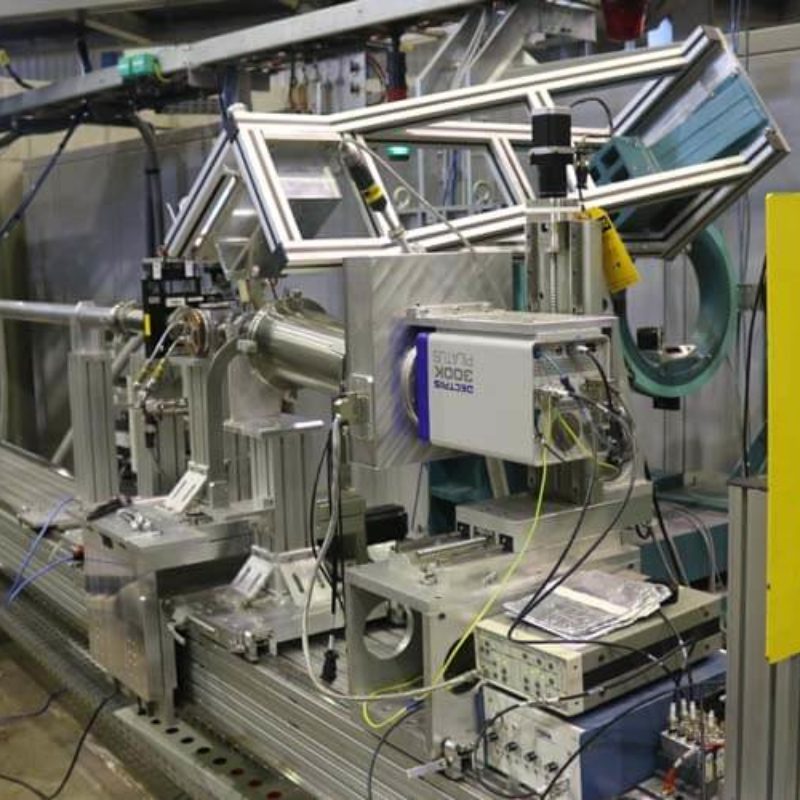
Português:
Cabana experimental da Linha de luz SAXS1.
English:
SAXS1 beamline experimental hutch.
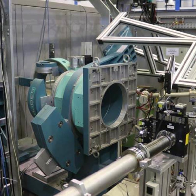
Português:
Goniômetro usado como suporte de detector, para realização de WAXS.
English:
Goniometer used for detector support, for the pourpose of doing WAXS.
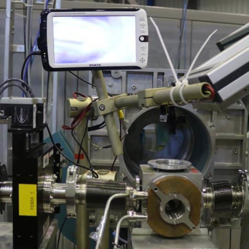
Português:
Câmara de porta-amostra, com controle de temperatura por banho térmico e visualização da amostra por câmera.
English:
Sample holder chamber, with thermal bath temperature control and sample visualized by camera.
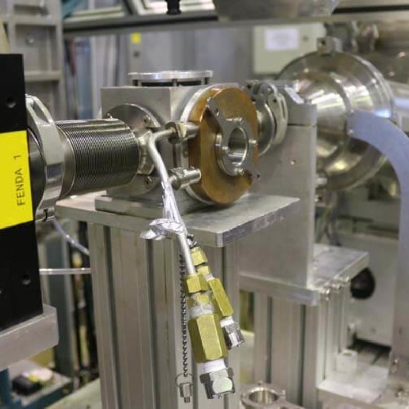
Português:
Vista lateral do porta-amostra.
English:
Lateral view of the sample holder.
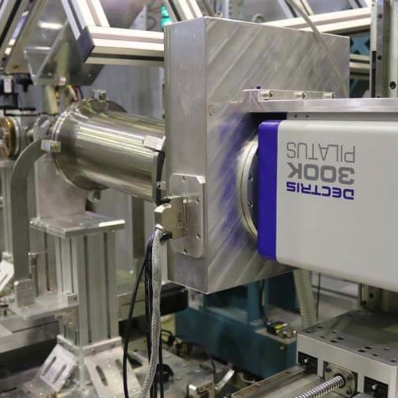
Português:
Detector Pilatus 300K da Dectris, usado para aquisição de imagens de SAXS.
English:
Pilatus 300K detector, from Dectris, for acquisition of SAXS images.
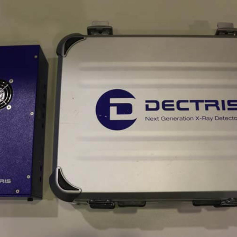
Português:
Detector Pilatus 100K da Dectris, usado para aquisição de imagens de WAXS.
English:
Pilatus 100K detector, from Dectris, for acquisition of WAXS images.
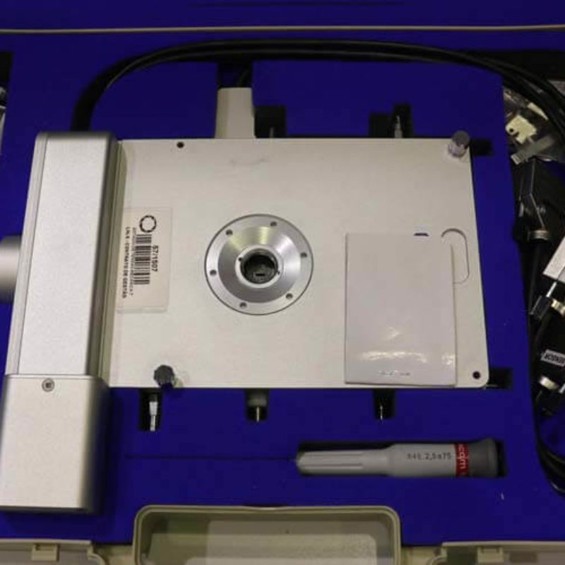
Português:
Porta-amostra TST350, da Linkam, para estudo de amostras sob tensão mecânica, com controle de temperatura (-196°C - 350°C).
English:
TST350 sample holder, from Linkam, for studies of samples under mechanical tensile, with temperature control (-196°C - 350°C).
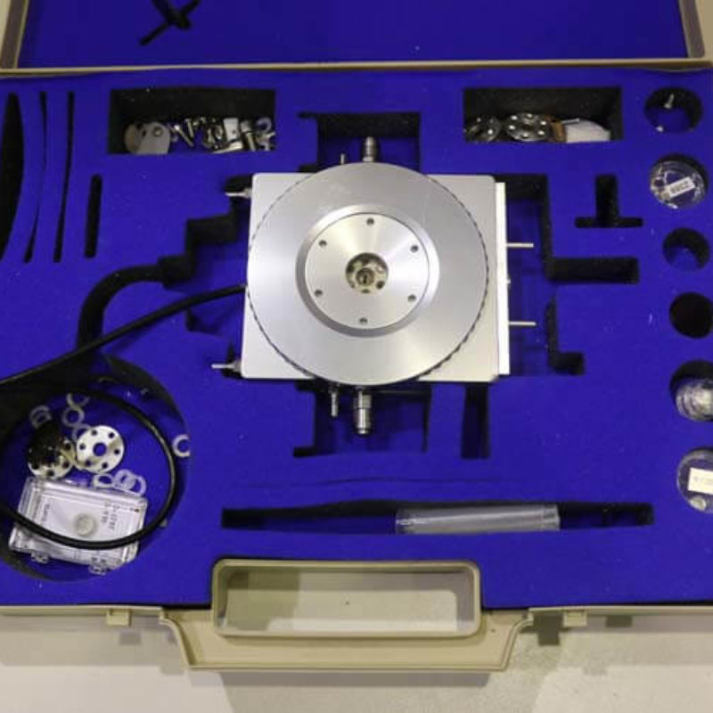
Português:
Porta-amostra DSC600, da Linkam, para estudo de amostras com controle de temperatura (-196°C - 600°C).
English:
DSC600 sample holder, from Linkam, for studies of samples with temperature control (-196°C - 600°C).
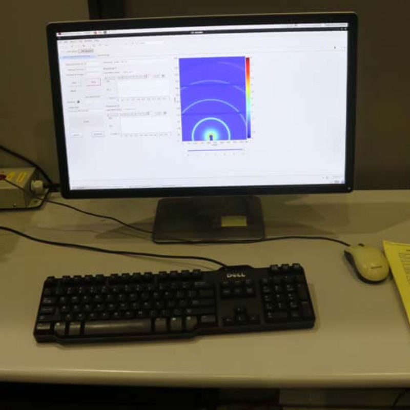
Português:
Programa de controle para realização dos experimentos de SAXS e WAXS.
English:
Control software for doing SAXS and WAXS experiments.
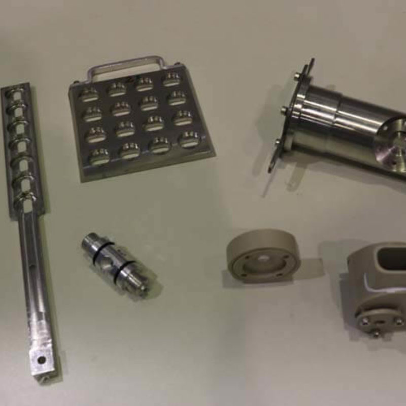
Português:
Diferentes tipos de porta-amostras personalizados da linha de luz SAXS1, para diversos tipos de amostras, como sólidos, líquidos e géis.
English:
Different type of sample holder developed in-house of SAXS1 beamline, for several types of samples, as solids, liquids and gels.
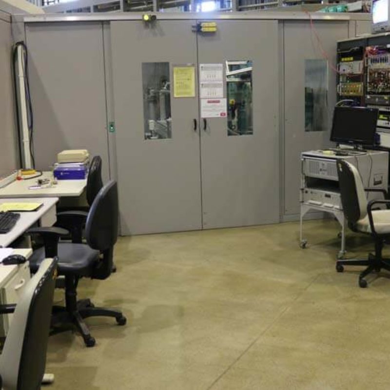
Português:
Estação de controle e preparo de amostra da linha de luz SAXS1.
English:
Control station and sample preparation of SAXS1 beamline.