CONTACT & STAFF
For more information on this beamline, contact us.


The XAFS2 beamline is an experimental station dedicated to X-ray Absorption Spectroscopy in the hard x-rays energy range (3.5 to 17.0 keV). It focus on the study of the atomic-level structure as well as in the electronic and magnetic properties of matter, with applications in a wide range of scientific fields, such as atomic and molecular physics, chemistry, biology, environmental and geosciences and cultural heritage. Experimental techniques available include Fluorescence Spectroscopy, X-ray Excited Optical Luminescence, X-ray Reflectivity and Combined X-ray Absorption Fine Structure and X-Ray Diffraction.
The XAFS2 is a general-purpose X-ray absorption beamline. Since the completion of the commissioning works in 2007, a large number of users have been using this experimental facility in order to perform several kinds of experiments in different scientific areas. After approximately 7 years in operation this beamline has been substantially updated in order to improve its experimental possibilities. A 4-circle Huber diffractometer has been recently incorporated to perform combined experiments. This equipment collects XRD patterns with the XAFS.
Through the development of a new sample environment, it is now also possible to perform these measurements in situ/operando conditions. Other upgrades include a complete remodeling of the beamline software and its control system. The control system of the beamline has been renovated by the installation of a PXI. The PXI is from National Instruments (PXI-NI) and communicates with Galil/Parker controllers on an EPICS platform. Some parts of the motors were changed in order to improve performance with the upgrade and there were also important changes to the control hardware. The Windows operational system was replaced with Red Hat Linux and the 3-WinDCM control system with EPICS. Furthermore, a new python based script (Py4Syn) was added. This provides high-level abstraction for device manipulation, scan routines, real-time plots and more. This package was created with the aim of providing a simple yet powerful tool to allow scientists and users to develop their own scripts for data acquisition. For user-friendly interface builds a Control System Studio (CS-Studio) is used. The next step with the XAFS2 upgrade, namely, towards a high-throughput XAFS beamline, will involve testing the viability of performing QEXAFS.
For more information on this beamline, contact us.
The following experimental techniques and setups are available to users in this beamline. To learn more about the techniques’ limitations and requirements (sample, environment, etc.) contact the beamline coordinator before submitting your proposal.
X-ray absorption spectroscopy (XAS) is a widely used technique for determining the local geometric and/or electronic structure of matter. This setup is optimized for transmission and fluorescence XAS on “standard samples” in standard sample holders. The setup for these experiments has three ionization chambers, a multi-element solid state Ge fluorescence detector and a 5K cryostat. In general, the samples for these environments are membranes, pellets, thin films, liquid and bulk. More attention on sample homogeneity preparation must be given when in transmission mode.
Recent publications in this setup:
Sampaio DV, Souza NRS, Santos JCA, Silva DC, Fonseca EJS, Kucera C, Faugas B, Ballato J, Silva RS; Translucent and persistent luminescent SrAl2O4:Eu2+Dy3+ ceramics. CERAMICS INTERNATIONAL 42, 4306 (2016). doi:10.1016/j.ceramint.2015.11.108
Cappellari P.S., Buceta D., Morales G.M., Barbero C.A., Moreno M.S., Giovanetti L.J., Ramallo Lopez J.M., Requejo F.G., Craievich A.F., Planes G.A.. Synthesis of ultra-small cysteine-capped gold nanoparticles by pH switching of the Au(I)–cysteine polymer. JOURNAL OF COLLOID AND INTERFACE SCIENCE 441, 17 (2015). doi:10.1016/j.jcis.2014.11.016
This setup allow to submit the samples to different gas atmospheres, while heating up to 1000°C and work on transmission or fluorescence mode. There is a tubular furnace used in transmission and the samples are prepared as pellets. For in-situ fluorescence the sample are powder diluted in boron nitride inside a capillary. The optical table currently has three ionization chambers, a set of slits and motorized stages for user-supplied equipment. If you seek to use the in-situ setup you should contact us well in advance of any proposal deadline to establish technical feasibility.
Recent publications in this setup:
Coletta V.C.; Marcos F.C.F.; Nogueira F.G.E., Bernardi M.I.B; Michalowicz A.; Goncalves R.V.; Assaf, E.M.; Mastelaro V.R.. In situ study of copper reduction in SrTi1-xCuxO3 nanoparticles. PHYSICAL CHEMISTRY CHEMICAL PHYSICS 18, 2070 (2016). doi: 10.1039/C5CP05939A
Ribeiro R.U., Meira D.M., Oliveira D.C., Rodella C.B., Bueno J.M.C., Zanchet D. Probing the stability of Pt nanoparticles on encapsulated in sol-gel Al2O3 using in situ and ex situ characterization techniques. APPLIED CATALYSIS A-GENERAL 485, 108 (2014). doi:10.1016/j.apcata.2014.07.039
This setup allows to get information about the optical behavior of a material when it irradiated with X-rays. It is possible to measure the emission spectra and/or the integrated luminescence as an energy function. Low temperature measurements (cryo-XEOL) are available using a cryostat, reaching 15K. If you seek to use the XEOL setup you should contact us well in advance of any proposal deadline to establish technical feasibility.
Recent publications in this setup:
Hora DA, Andrade AB, Ferreira NS, Teixeira VC, Rezende, MVS (2016). X-ray excited optical luminescence of Eu-doped YAG nanophosphors produced via glucose sol–gel route. Ceramics International, 42(8), 10516-10519 (2016).
doi: 10.1016/j.ceramint.2016.03.142Rezende M.V., Montes P.J.R., Andrade A.B., Macedo Z.S., Valerio M.E.G.. Mechanism of X-ray excited optical luminescence (XEOL) in Europium doped BaAl2O4 phosphor. Physical Chemistry Chemical Physics, 18, 17646-17654 (2016). doi: 10.1039/C6CP01183G
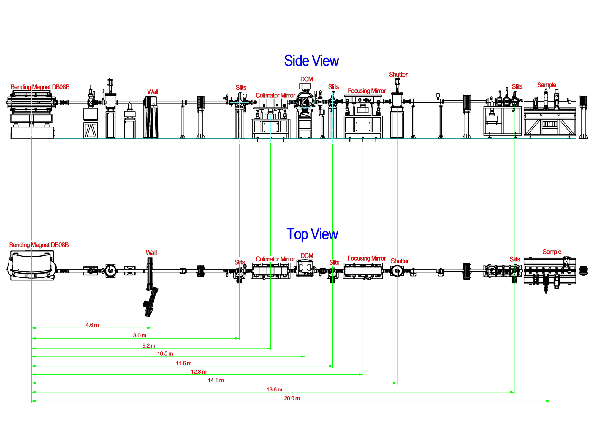
| Element | Type | Position [m] | Description |
|---|---|---|---|
| Source | Bending Magnet | 0.0 | Bending Magnet D08 exit B (15°) |
| 1st cooled-slit system | Two cooled UHV slit systems – vertically and horizontally – based on four independent mechanical feedthroughs, each one supporting at its ends a Ta blade mounted on a copper block. | 8.0 | Defines the lateral and vertical dimensions of the polychromatic beam impinging on the first mirror |
| 1st mirror | Rh-coated cylindrical vertical collimating mirror with a ~3mrad grazing incidence angle | 9.2 | Collimate vertically the white radiation and sends a parallel synchrotron beam onto the two flat Si(111) Double Crystal Monochromator (DCM) |
| Double Crystal Monochromator (DCM) | Flat Si(111) Double Crystal Monochromator | 10.5 | The DCM has fixed exit geometry and is the only optical element with thermal stabilization (i.e. water cooled). |
| 2nd slit system | Two UHV slit systems – vertically and horizontally – based on four independent mechanical feedthroughs, each one supporting at its ends a Ta blade mounted on a copper block. | 11.6 | Without refrigeration, these slits are used to eliminate the spurious background radiation. |
| 2nd mirror | Rh-coated toroidal bendable mirror | 12.8 | It refocuses vertically and horizontally the monochromatic beam of approximately 450 µm in diameter at the sample position |
| Parameter | Value | Condition |
|---|---|---|
| Energy range [keV] | 3.5 – 17.0 | Si(111) |
| Energy resolution [ΔE/E] | 1.7 x 10⁻⁴ | at 7 keV |
| Beam size [µm2, FWHM] | 450 x 250 | at sample position |
| Flux density at sample [ph/s/mm2] | 2.78 x 10⁹ photons/s at 7 keV and 100mA | Measured with a photodiode 100% efficient |
| Harmonic Content | 3.94 x 10⁻⁵ | at 7.5 keV |
| Instrument | Type | Model | Manufacturer | Specifications |
|---|---|---|---|---|
| Detector | Ion Chamber | – | Two electrodes in a distance of 14 mm. Length of 137 mm, 221 mm and 381 mm. A 25 µm kapton window with a in/out area of 12 x 30 mm2 | LNLS in-house development |
| Detector | 15-element Germanium Solid State Detector (SSD) | G-15 SSD | High counting rate capability – 300.000cts/s. Si detector total active area – 750 mm2. Element sensitive thickness – 5 mm. N2 liquid cooled. | Canberra |
| Detector | Electron Detector | – | Collector electrode (He medium) in an electric field produced by a 60V battery. The output is a signal amplification of about two orders of magnitude | LNLS in-house development |
| Detector | Photomultiplier | Model R928 | Side-on; V = -1200 V. It output is in current mode. | Hamamatsu |
| Detector | Spectrometer | USB2000+ | covers the 200-1100 nm range and connects to light sources, optical fibers and other accessories | Ocean Optics |
| Furnace | Transmission/ Fluorescence | Capilar | Max Temp.: 900 C, Temp Rate: 10C/s. E5CK-T Ramp/Soak Process Controller-Omron. Sample holder for capillars (ID 0.8 mm/ OD 1mm) (ID 1 mm/ OD 1.2 mm) (ID 2 mm/ OD 2.2 mm) | LNLS in-house development |
| Furnace | Transmission | Tubular | Max Temp.: 1100 C, Temp Rate: 10C/s. E5CK-T Ramp/Soak Process Controller-Omron. Sample holders of 8 mm and 4 mm diameters | LNLS in-house development |
| Diffractometer | 4-circle | 424-511.1 | Resolution (θ, 2θ, φ, χ) = 0.001° | Huber |
| Mass spectrometer | Gas Analysis System | OmniStar QMS 200 | Tungsten (standard) filament. Mass range 1-100 amu. Gas flow rate 1-2 sccm. Qualitative and quantitative gas analysis. | Pfeiffer Vacuum |
| Cryostat | Cryogen free and top loading sample in helium, fast sample change. | Omniplex: CS204*F-FMX-19OP | High cooling power and fast cooldown. 4 K cold head (0.2 W at 4.2 K). 180° Kapton window for x-ray fluorescence detection mode | ARS |
| Cryostat for cryo-XEOL | Cooling power He closed cycle cryocooler | DE-202 Cryocooler series | Tmin = 15K |
All beamline controls are done through EPICS (Experimental Physics and Industrial Control System), running on a PXI from National Instruments. The data acquisition is done using a Red Hat workstation with the Py4Syn, developed at LNLS by SOL group. CSS (Control System Studio) is used as a graphical interface to display and control the beamline devices.
The beamline can be operated remotely by using LabWeb for experiments in conventional transmission (without furnaces and gases). We are working to improve these possibilities on Sirius. For more details, contact the beamline coordinator.
Click here to download the XAFS2 beamline manual (in portuguese).
Users are required to acknowledge the use of LNLS facilities in any paper, conference presentation, thesis and any other published material that uses data obtained in the execution of their proposal.
FIGUEROA, S. J. A.; MAURICIO, J. C.; MURARI, J.; BENIZ, D. B.; PITON, J. R.; SLEPICKA, H. H.; FALCÃO DE SOUSA, M.; ESPÍNDOLA, A. M.; LEVINSKY, A. P. S.. Journal of Physics: Conference Series 712 (2016) 012022. DOI: 10.1088/1742-6596/712/1/012022
The XAFS2 is a general-purpose X-ray absorption beamline. It is the second one built at the LNLS. After approximately 7 years in operation this beamline has been substantially updated in order to improve its experimental possibilities. Recently arrived, a 4-circle Huber diffractometer has been incorporated to perform combined experiments. This collects XRD patterns with the XAFS. Through the development of a new sampling environment it is now also possible to perform these measurements in situ/operando conditions. Other upgrades include a complete remodelling of the beamline software and its control system. The following new systems are crucial for the next steps that are currently underway at the beamline, namely, (i) enabling remote access for users and (ii) the testing of QEXAFS measurements.
Scientific publications produced with data obtained at the facilities of this beamline, and published in journals indexed by the Web of Science, are listed below.
Chocobar-Ponce, S. ;Prado, C. ;Tabernero, R. ;Ilina, N. ;Pagano, E. ;Ramallo-López, J. M.;Mizrahi, M.;Rosa, M.. The reduction of Cr(VI) in Salvinia minima, possible involvement of an h-type thioredoxin, Environmental Science and Pollution Research, v.29, p. 3958–3966, 2022. DOI:10.1007/s11356-021-15967-z
Sampaio, D. V.;Pena, R. B. ;Moulton, B. J. A.;Rezende, M. V. dos S.;Silva, D. do C. ;Silva, R. S. da;Cunha, T. R. da;Mastelaro, V. R.;Zanotto, E. D.;Pizani, P. S.. Chromium in lead metasilicate glass: Solubility, valence, and local environment via multiple spectroscopy, Ceramics International, v.48, n.1, p.173-178, 2022. DOI:10.1016/j.ceramint.2021.08.377
Teixeira, M. M. ;Santos, L. C. ;Tello, A. C. M. ;Almeida, P. B. ;Silva, J. S. da ;Laier, L. O. ;Gracia, L.;Teodoro, M. D.;Silva, L. F. da;Andrés, J.;Longo, E.. aeAg2WO4 under microwave, electron beam and femtosecond laser irradiations: Unveiling the relationship between morphology and photoluminescence emissions, Journal of Alloys and Compounds, v.903, p.163840, 2022. DOI:10.1016/j.jallcom.2022.163840
Marcos, F. C. F.;Cavalcanti, F. M. ;Petrolini, D. D.;Lin, L. ;Betancourt, L. E. ;Senanayake, S. D. ;Rodriguez, J. A.;Assaf, J. M.;Giudici, R. ;Assaf, E. M.. Effect of operating parameters on H2/CO2 conversion to methanol over Cu-Zn oxide supported on ZrO2 polymorph catalysts: Characterization and kinetics, Chemical Engineering Journal, v.427, p.130947, 2022. DOI:10.1016/j.cej.2021.130947
Gomes, F. P. ;Barreto, M. S. C.;Amoozegar, A. ;Alleoni, L. R. F.. Immobilization of lead by amendments in a mine-waste impacted soil: Assessing Pb retention with desorption kinetic, sequential extraction and XANES spectroscopy, Science of the Total Environment, v.807, n.1, p.150711, 2022. DOI:10.1016/j.scitotenv.2021.150711
Matte, L. P.;Thill, A. S.;Lobato, F. O.;Novôa, M. T. ;Muniz, A. R. ;Poletto, F. S.;Bernardi, F.. Reduction-Driven 3D to 2D Transformation of Cu Nanoparticles, Small, v.18, n.7, p.2106583, 2022. DOI:10.1002/smll.202106583
Bravo, C. A. O.;Figueroa, S. J. A.;Portela, R. ;Chagas, C. A.;Bañares, M. A. ;Toniolo, F. S.. Elucidating the structure of the W and Mn sites on the Mn-Na2WO4/SiO2 catalyst for the oxidative coupling of methane (OCM) at real reaction temperatures, Journal of Catalysis, v.408, p.423-435, 2022. DOI:10.1016/j.jcat.2021.06.021
Português:
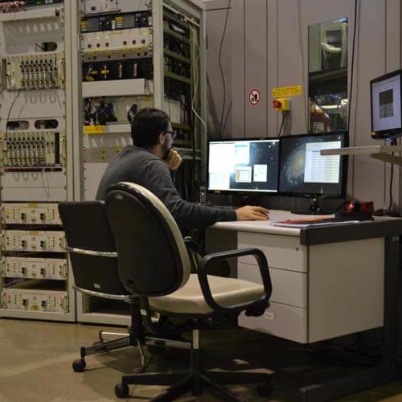
Processo de aquisição de dados na Linha de Luz XAFS2.
English:
Data acquisition process at the XAFS2 beamline.
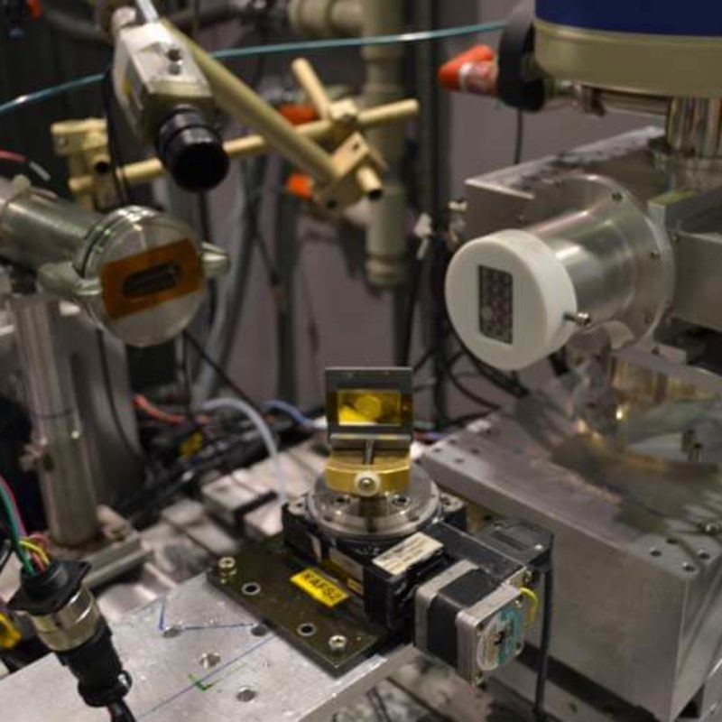
Português:
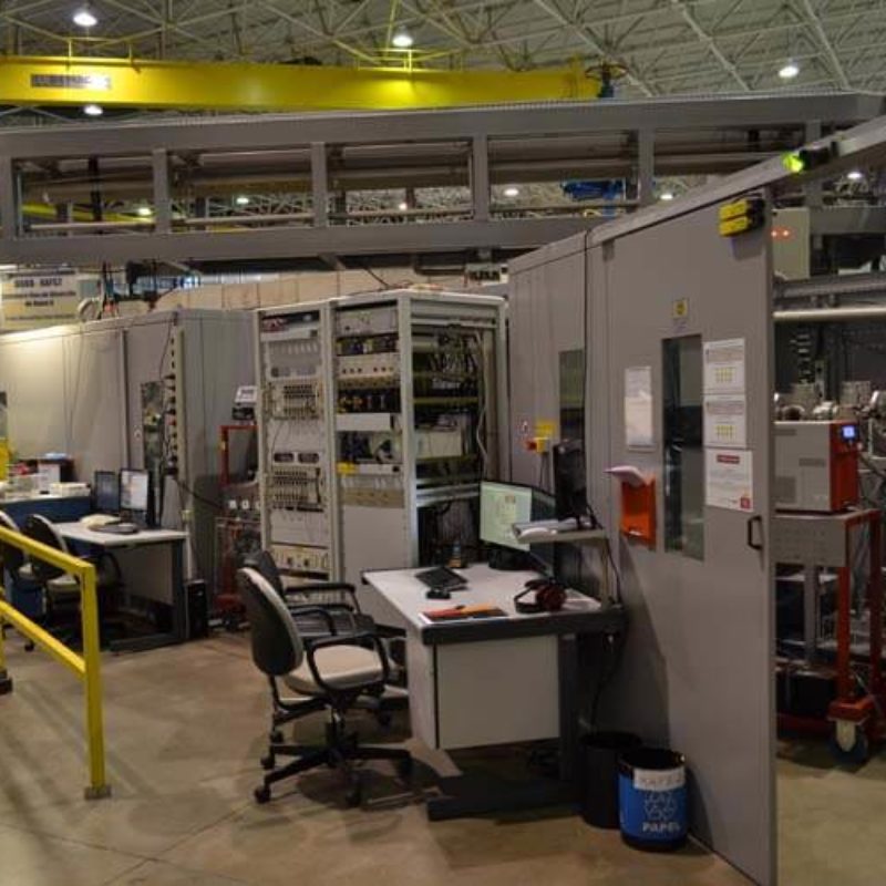
Visão geral das estações da Linha de Luz XAFS2.
English:
Overview of the XAFS2 beamline stations.
Português:
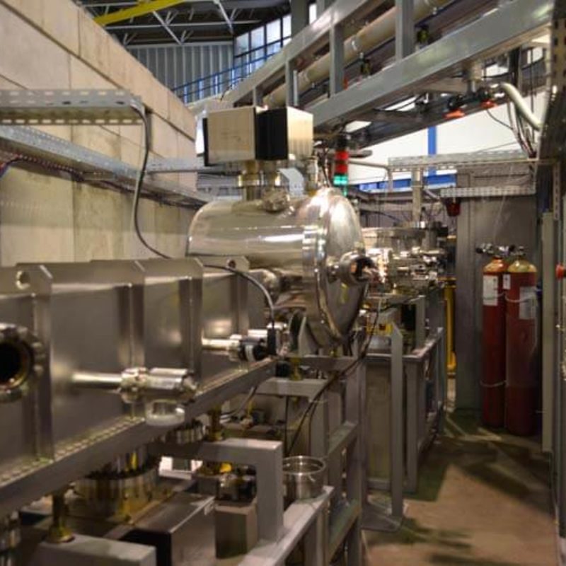
VIsão geral da Cabana Óptica. Da esquerda para direita: primeira câmara de espelho (forma retangular), câmara do monocromador DCM (forma cilíndrica) e segunda câmara de espelho (forma retangular).
English:
Overview of the optical hutch. From left to right: first mirror chamber (rectangular shape), DCM monochromator chamber (cylindrical shape) and second mirror chamber (rectangular shape).
Português:
Visão Monochomador de Cristal Duplo. Motores de picômetro controlam rotações nos três eixos e são mostrados na parte superior do segundo cristal. Água é usada para manter uma temperatura constante de 24ºC no primeiro cristal.
English:
Close view of the Double Crystal Monochromator. Picometer motors that control the pitch, roll and yaw displacements are shown at the top of the second crystal. Water is used to maintain a fixed temperature of 24ºC in the first crystal.
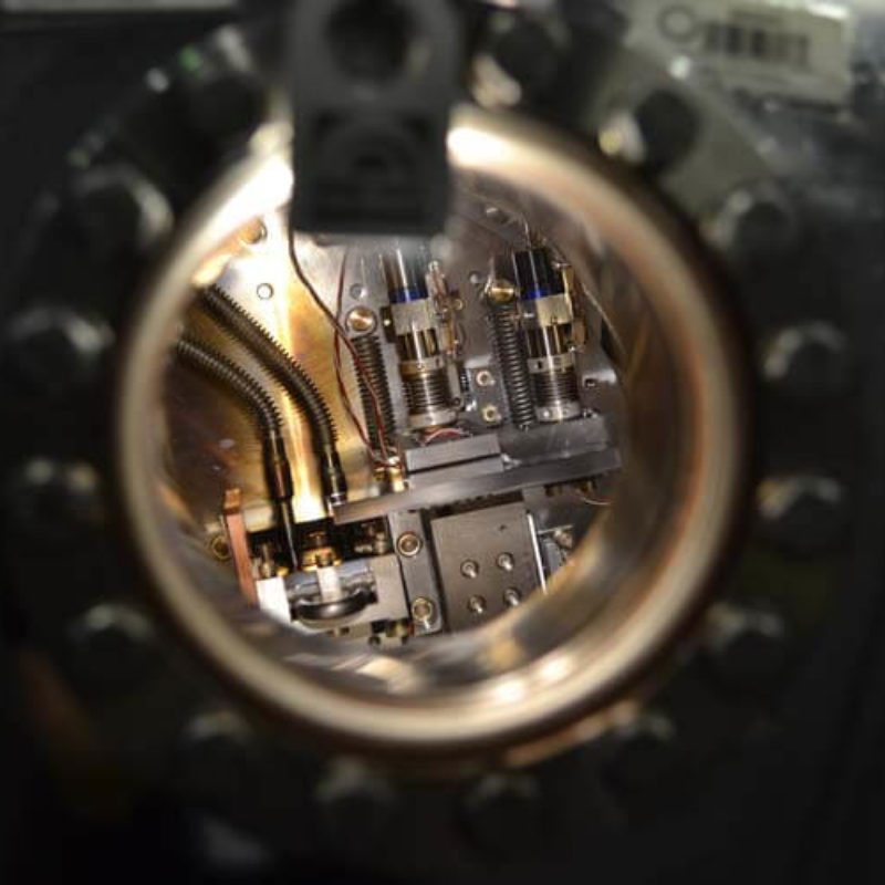
Português:
Visão detalhada da configuração para medições de fluorescência na Linha de Luz XAFS2. Um goniômetro (em marrom) ajuda a posicionar a amostra com o melhor ângulo para maximizar o sinal de fluorescência. Um detector Canberra Ge-15 elementos é usado para obter o sinal de fluorescência.
English:
A detailed view of the setup for fluorescence measurements at XAFS2 beamline. A goniometer (in brown) helps placing the sample attached on a slide at the best angle in order to maximize the fluorescent signal. A Ge-15 elements Canberra detector is used to collect the fluorescence signal.