

Study shows the validity of mini-brains as a model for embryonic neurodevelopment.
The study of the embryonic development of the human brain and the distribution of nutrients during this phase was for a long time limited to animal models and dead human tissues. Much has changed with the so-called organoids: mini-organs created in vitro in the laboratory, from a few cells of the organ themselves, from embryonic stem cells or from pluripotent cells. These organoids present a three-dimensional micro-anatomy similar to the real organ but more simplified.
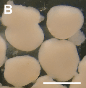
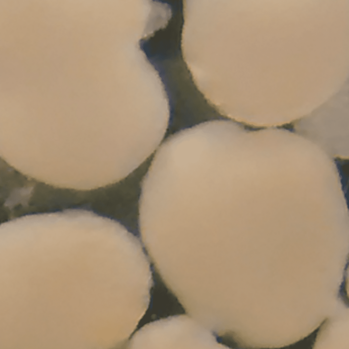
Figure 1: Mini-brain with 45 days. The white line defines a scale of 1 mm.
Understanding the distribution of chemical elements that are essential to the embryonic brain can elucidate, for example, the mechanisms behind a variety diseases related to the neuronal development, including those manifested in adulthood.
Recently, a group of researchers from the D’Or Institute for Research and Education, the Federal University of Rio de Janeiro, the Federal Institute of Education, Science and Technology of Rio de Janeiro, and the Brazilian Synchrotron Light Laboratory, used the LNLS facilities to study the distribution of chemical elements in human brain organoids, also called mini-brains.
Researchers used the XRF X-Ray Fluorescence beamline to produce a map of the chemical elements present in the mini-brains, such as phosphorus, sulfur, potassium, calcium, iron, zinc and others. This is possible because, when atoms in a sample are excited with X-Rays, each chemical element re-emits radiation in a specific way, producing a unique signature that allows its identification.
Organoids were studied in two phases, equivalent to different moments of embryonic development: one in which there is intense cellular multiplication and another in which there is the formation of structures between neurons. It was observed that the distribution of elements is modified between the phases, indicating either the operation of specific receptors for certain chemical elements or a specific use of these elements in the organoid specific cellular environment.
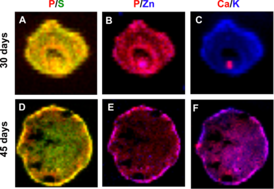
Figure 2: Maps of the concentration of different elements in cerebral organoids at 30 (top) and 45 days (bottom) of development, for pairs of elements: Phosphorus / Sulfur (A) and (D); Phosphorus / Zinc (B) and (E); and, Potassium / Calcium (C) and (F).
The results show the potential of using mini-brains, allied to X-Ray Fluorescence techniques, as a model for exploring changes in micronutrient distribution during brain development.
Source: Sartore RC, Cardoso SC, Lages YVM, Paraguassu JM, Stelling MP, Madeiro da Costa RF, Guimaraes MZ, Pérez CA, Rehen SK. (2017) Trace elements during primordial plexiform network formation in human cerebral organoids. PeerJ 5:e2927. DOI: 10.7717/peerj.2927
A detailed characterization of platinum-cerium-alumina model catalysts under Water Gas Shift conditions have been performed.
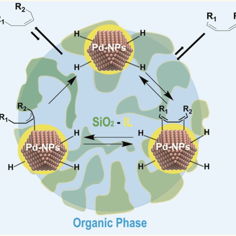
Results suggest that ionic liquid hybrid organosilica/Pd-NPs under multiphase conditions operate akin to catalytically active membranes.