

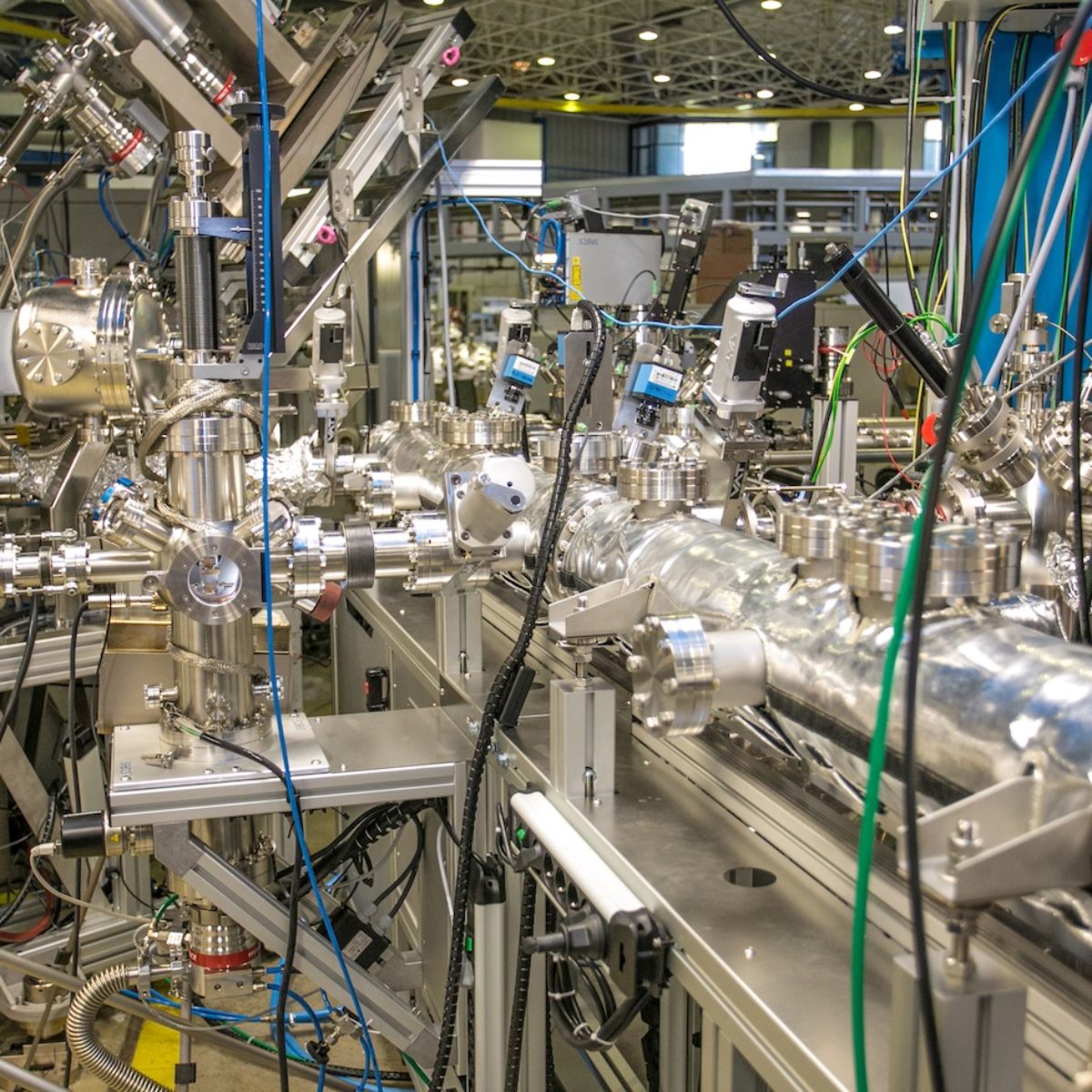
ARPES (angle-resolved photoemission spectroscopy experiments) station installed at PGM beamline, for UV and soft X-ray spectroscopy
New photoelectron spectroscopy station is suitable for analysis of the electronic structure of materials
The PGM beamline for UV and soft X-ray spectroscopy has a new photoelectron spectroscopy end station, suitable for analysis of the electronic structure of materials. Researchers at the UVX light source have already used this new instrumentation, which will be transferred to the Sirius light source when it starts operation.
The new station, built for the study of surface science, consists of an electron analyser suitable for angle-resolved photoemission spectroscopy experiments (ARPES) and a flexible system for growth and characterization of samples in situ. According to Júlio Criginski Cezar, researcher and PGM beamline coordinator, this instrumentation coupled to a synchrotron light source composes a unique experimental setup.
One of the main purposes of this experimental station is to promote the “layer by layer growth”: technique based on the deposition of materials onto a substrate as thin films, usually by evaporation. This process is widely used for the development of new technologies and products in microelectronics, radiofrequency materials, optical components, protective coatings for mechanical parts, catalysis, among others.
The development of increasingly complex thin films opens new possibilities for the modification and control of material properties. Heterostructure-based films, for example, consisting of very thin layers of different materials with different structures, can exhibit novel properties not found on the materials in their isolated form.
Equipment
The core of the ARPES station is a SPECS analyzer Phoibos 150-CCD, coupled to a high precision six degrees of freedom manipulator, allowing measures from 12 to 400 K. The analyzer is connect through a modular ultra-high-vacuum transfer system to several instruments used to grown and pre-characterize the samples. Films can be grown within an MBE (Molecular Beam Epitaxy) growth chamber, equipped with Low Energy Electron Diffraction (LEED) and Reflectivity High Energy Electron Diffraction (RHEED), or in the laser ablation system (PLD, Pulsed Laser Deposition), which is under commissioning. Pre characterization of the films can be done using a scanning microscope that allows simultaneous acquisition in STM (Scanning Tunneling Microscopy) and AFM (Atomic Force Microscopy) modes.
In addition, it is available for the first time at the station a Photoemission Electron Microscope (PEEM) that allows obtaining spectroscopic information with submicron spatial resolution.
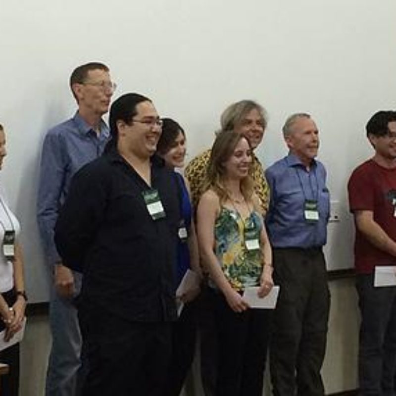
Jessica Oliveira and Willian Takemitsu had their posters awarded during the AutoOrg Conference
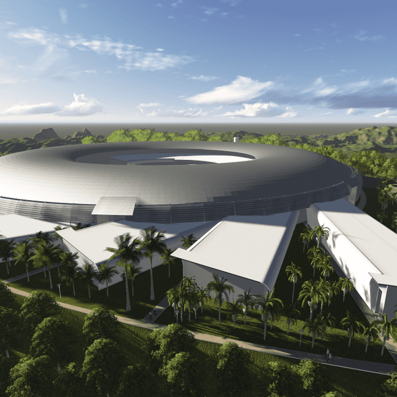
FAPESP and FINEP are selecting proposals of companies that are interested in developing components for the new light source