

Visualization of the polymeric matrix of the hollow membranes analyzed at the Cateretê beamline
Article published in Communications Materials presents significant findings and discusses the possibilities offered by this technique combined with synchrotron light sources
Porous materials play key roles in a variety of contexts, from transporting water and nutrients in biological systems to storing oil and water in reservoirs of rock. And synthetic polymer membranes are essential to separation processes, as in the case of chromatography.
They have well-established applications in water desalination, hemodialysis, and gas separation, and these uses are expanding into processes that filter out pollutants from contaminated water. Their benefits include energy efficiency, smaller carbon footprint, and compact design that provides a large area of membrane in a small volume.
Membranes that can fulfill technological objectives have complex porous structures that ensure the required selectivity, mechanical stability, and characteristics for rapid transport; the effectiveness and performance of these membranes is defined by characteristics such as porosity and interconnectivity, which can be particularly difficult to measure when they are brought down to the nano scale.
Techniques such as scanning electron microscopy (SEM), atomic force microscopy (AFM), and transmission electron microscopy (TEM) have helped scientists better understand the transport mechanisms in these applications and develop membranes for different purposes. But even though they are powerful, these techniques also have significant limitations: the samples must be dehydrated and covered with a metallic layer, and must also remain in a vacuum during analysis, which can affect the structure of these membranes and hinder analysis under near-real conditions.
Furthermore, when the pores of the material reach the nano scale, the total sample volume must be significantly reduced in order to attain the resolution necessary for analysis. Within this context, X-ray tomography has emerged as a good alternative. This method not only offers non-destructive visualization, but samples can be analyzed in ambient conditions with significantly larger total sample volumes.
Conventional X-ray tomography, which analyzes different absorption in different parts of a sample, faces challenges related to resolution limits when analyzing less dense materials (such as membranes). But as new fourth-generation synchrotron light sources have recently come online and ptychographic X-ray tomography has been developed, images of these materials can be obtained with nanometric resolution.
Ptychographic X-ray computed tomography (PXCT) is a powerful phase-contrast imaging technique that uses a series of two-dimensional projections of the object from different angles to reconstruct its three-dimensional structure in high resolution, revealing information about porosities and interconnectivity.
3D visualization of the pore interconnectivity
The article published in the journal Communications Materials by a group of researchers from the Brazilian Center for Research in Energy and Materials (CNPEM) and King Abdullah University of Science and Technology (KAUST) highlights the capacities of PXCT and describes work carried out to
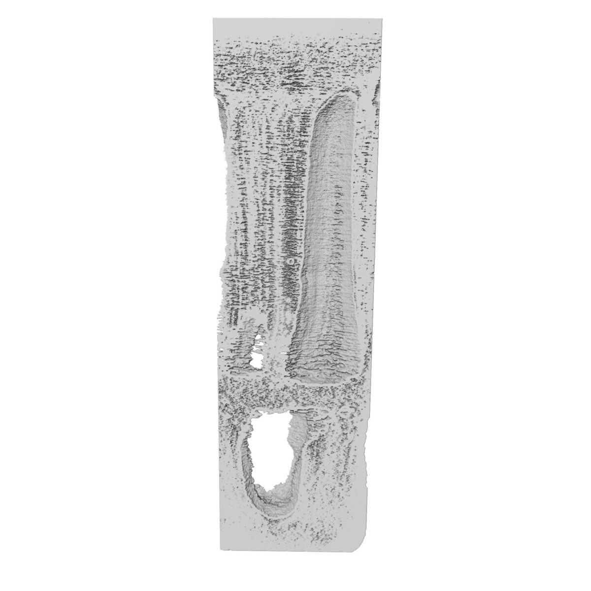
obtain 3D images with spatial resolution of 26 nanometers, the highest for porous polymer materials so far.
“This type of experiment promotes the construction of a ptychography nanotomography community in Latin America, and offers new opportunities for experiments for those who work with polymeric structures in different areas of application. This broad view makes it possible for you to have data from different size scales in a single experiment, which is generally done by combining various different techniques,” notes Carla Pólo, a CNPEM researcher and coauthor of the article.
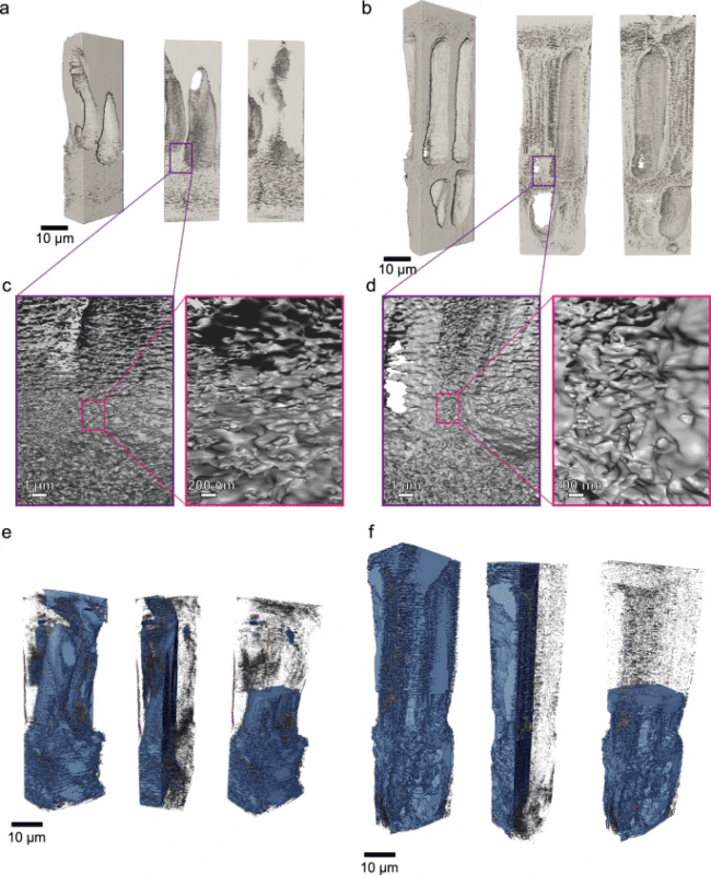
To achieve this landmark, the scientists prepared two hollow polymeric membranes using polyethylenimine (PEI), diethylene glycol (DEG), N-methylpyrrolidone (NMP), and bore liquid (a coagulant) containing two different compositions.
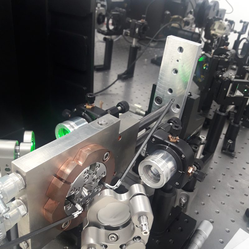
The analysis was conducted using an X-ray beam produced by Sirius’s Cateretê beamline. Several diffraction patterns were collected for each of the samples at different points and projection angles. By processing the massive quantity of data collected, three-dimensional images of the samples could be reconstructed at unprecedented resolution levels.
The 23 nm x 23 nm x 23 nm spatial resolution allowed the researchers to extract quantitative data about the material and revealed the existence of a dense layer on the external surface of the membrane with less than 5% porosity, while the inside of the membrane exhibited a spongy structure with porosity of up to 74% in some areas.
The analysis also extended to the distribution of pore size within the membranes, a crucial characteristic that influences membrane performance in practical applications such as water filtration or gas separation.
The interconnectivity of pores throughout the entire membrane could also be assessed; although most pores were connected to others, there were notable differences in the way they were distributed in the membranes made with different compositions. This affected the performance of the membranes, impacting their capacity to allow substances to pass through and their selectivity, emphasizing the importance of understanding the structure to improve its practical applicability.
As noted in the article’s conclusion, the highly coherent X-ray beam produced by 4th generation synchrotron light sources like Sirius allow non-destructive analyses through PXCT on the nanometric scale. And these analyses are not limited to measurements of porous structures: they can also be applied to investigations of other soft-material nanostructures.
“Ptychographic X-ray tomography complements conventional electron microscopy imaging techniques and creates new possibilities for research and development of porous materials, contributing to progress in various areas ranging from treating polluted water to biomedicine and providing a deeper understanding of the structure and functionality of these complex materials,” concludes CNPEM researcher and article coauthor Florian Meneau.
The research used diamond anvil cells (DAC) together with synchrotron light to investigate superconductors under extreme pressures
Review article was highlighted in the Applied Physics Reviews journal and explains how computed micro- and nanotomography can be used in fourth-generation synchrotrons like Sirius