

This support laboratory allows the preparation of advanced samples and their characterization using electron and optical microscopy techniques, complementary to the experiments in the beamlines
In addition to enabling extremely advanced experiments, the Sirius synchrotron light source, from the Brazilian Center for Research in Energy and Materials (CNPEM), a private non-profit organization under the supervision of the Brazilian Ministry of Science, Technology and Innovations (MCTI), aims also to provide all the necessary infrastructure for researchers to carry out their investigations.
As such, support laboratories installed around the beamlines will meet the demands of users regarding the preparation and conditioning of samples, carrying out controlled chemical reactions and the use of equipment that may be unavailable at the researcher’s institution.
Microscopic Samples Laboratory
This is the case of the Microscopic Samples Laboratory (LAM), whose main purpose is the preparation of advanced samples and their characterization using electron and optical microscopy techniques, complementary to the experiments in the beamlines.
In terms of advanced sample preparation, a modern SEM/FIB (Scanning Electron Microscope with Focused Ion Beam) dual beam system is used as a standard method, achieving high quality at the nanometric level. No other mechanical method is capable of preparing samples with this quality, especially from heterogeneous and hierarchical materials, when it is necessary to select specific regions for preparation. It enables in-situ large area sample preparation and high-resolution imaging and analysis of areas hundreds of microns across to ensure the collection of relevant and representative results. The system will also allow the deposition of some materials (carbon and tungsten) as fiducial marks on the samples to facilitate the location of some specific regions that will later be analyzed in the beamlines. Furthermore, it is possible to pre-characterize samples using various techniques, such as EDS, EBSD and cathode-luminescence. This process is essential to allow the best use of the measurement time at the beamines, ensuring the data highest quality. In addition, these analyses will also serve as complementary measurements to the X-ray experiments, increasing the robustness of the characterization that will be carried out later at the beamlines.
The facility also houses appropriate tools and equipment for the process of preparing nanometric samples and van der Waals heterostructures, as well as the optical characterization of different types of samples. This facility allows beamline users to prepare samples from one or a few atomic layers suitable for the nanoprobe technique. It is known that these materials when combined in van der Waals heterostructures form new patterns and present new physical properties different from the isolated material, such as superconductivity. Consequently, techniques with nanometric beams meet the need of researchers to respond to the effects of reduced dimensionality, inherent to the crystallographic level, on their electronic and optical properties. Therefore, its structure, defects, impurities and related optical phenomena can be explored by several of the Sirius beamlines, such as CARNAÚBA, IPÊ, SAPE, IMBUIA and EMA.
Hence, LAM will serve the entire Sirius community to manufacture high-quality, ultra-thin samples from multiple materials, as well as characterize and analyze the samples before or after the beamline experiments.
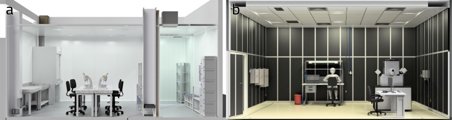
Figure 1: LAM layout: (a) FIB and (b) optical microscope areas.
Assembly and Commissioning
LAM facilities are designed to meet the acoustic, HVAC, utilities and control prerequisites needed to receive and operate the FIB with excellent reliability and performance.
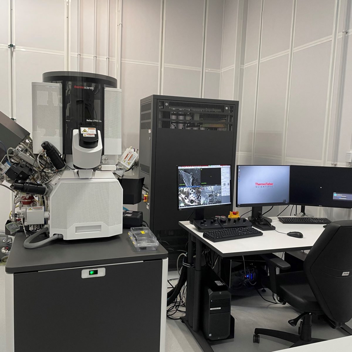
The equipment chosen to be installed in LAM is the Thermo Scientific Helios Plasma FIB CXe (PFIB) DualBeam, shown in Figure 2.
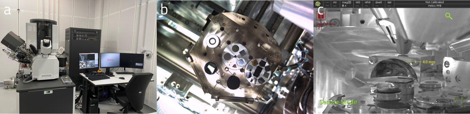
Figure 2: (a) PFIB in April 2022; (b) Specimen stage image obtained by In-Chamber Nav-Cam, (c) Specimen stage image obtained by an IR-CCD camera, showing the position of the sample with respect to electron and ion columns.
The laboratory was concluded in October 2021, and the PFIB installation process started in November 2021 and part of the installation was carried out until May 2022. The main components have already been installed: electron and ion gun; secondary electron detector (SE), allowing the acquisition of electronic images with a resolution of up to 0.6 nm; the gas injection system for deposition of materials (tungsten and carbon); and nano-manipulators for ultra-fine sample preparation and sequential cuts. Figure 3 shows some procedures performed on the PFIB during its installation.
Recently, the installation of the SPARC high-performance cathodoluminescence (CL) detection system is underway. The system is ideal for the collection and detection of cathodoluminescence emission, allowing fast and sensitive characterization of the materials at the nanoscale. SPARC is an upgradeable CL system suitable for nanophotonics and geological research. In addition to the CL system, other characterization techniques complementary to the experiments carried out on the beamlines will be installed soon: chemical and crystallographic analysis will be performed by Energy Dispersion X-Ray Spectroscopy (EDS) and Electron Backscatter Diffraction (EBSD), respectively.
The FIB will be available to users only at the end of the installation and commissioning phase, scheduled for the second half of 2022.
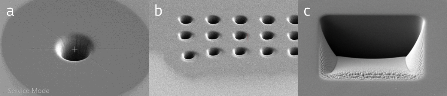
Figure 3: SE images of a) Individual micro-hole, b) micro-holes array and c) trenches in silicon substrate obtained by FIB milling.
In January 2022, the Nikon Eclipse LV100ND Optical Microscope and HQ Graphene 2D Transfer System were installed, as shown in Figure 4.
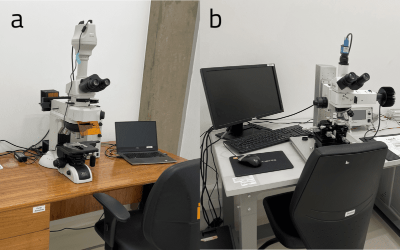
Figure 4: (a) Nikon Eclipse LV100ND Optical Microscope and (b) 2D Heterostructure Transfer System.
Specifically, Nikon’s optical microscope supports a wide range of observation methods: brightfield, darkfield, polarizing, and epifluorescence, which provides high-sensitivity detection of level differences and defects in samples, fluorescent properties, and high-contrast images. . Visualizing materials from atomic layers can be useful, and the fluorescence modality allows the identification of monolayers of some materials, separating them from the other layers, as shown in Figure 5.
Finally, the 2D Transfer System allows the fabrication of high quality van der Waals heterostructures. It is designed to accurately place a stamp on a substrate, which can be moved relative to each other in the x-, y- and z- directions and by tilting and rotation. This allows a high degree of freedom in the alignment of crystal flakes in the fabrication of twisted layers. The temperature (user defined, can be turned on or off) and vacuum of the substrate holder are touchscreen controlled. Thus, it is possible to build, for example, fully two-dimensional heterostructures with precise positioning and well-defined angles, as shown in Figure 5.
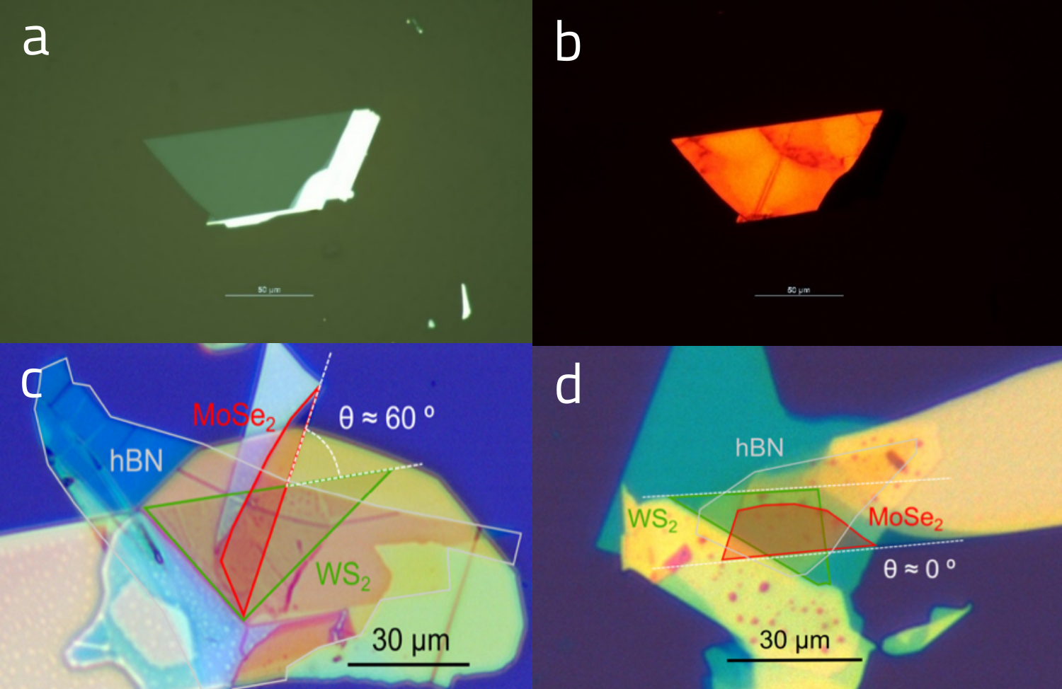
Figure 5: Two-dimensional crystal of WS2 exfoliated on a transparent membrane (a) optical microscope and (b) fluorescence image. Microscope image showing the fully two-dimensional van der Waals heterostructure of h-BN encapsulated WS2/MoSe2 monolayers with relative angle (θ) between layers of (c) θ = 0° and (d) θ = 60°.
The optical microscopy area was opened to internal and external users in March 2022. Proposals were received for the preparation of two-dimensional samples related to experiments on the EMA, IMBUIA and CARNAÚBA beamlines, and some of them have already been carried out.
Next Steps
The activities of the Microscopic Samples Laboratory’s team will continue with the completion FIB’s installation and the beginning of its technical and scientific commissioning. Additionally, although the optical microscopes are already operational, their installation was carried out in a temporary environment. Hence, the cleanroom in which this equipment will be definitively allocated will have its infrastructure project started.
New commissioning stage of Imbuia beamline allows the use of advanced infrared spectroscopy techniques at micro and nano scales
CARNAÚBA’s experimental station has detectors for simultaneous X-ray fluorescence, X-ray diffraction, X-ray excited optical luminescence and X-ray ptychography techniques, and is operating, during commissioning phase, from 6.4 keV to 14 keV