

CARNAÚBA’s experimental station has detectors for simultaneous X-ray fluorescence, X-ray diffraction, X-ray excited optical luminescence and X-ray ptychography techniques, and is operating, during commissioning phase, from 6.4 keV to 14 keV
CARNAÚBA is the longest beamline on Sirius, the new synchrotron light source from the Brazilian Center for Research in Energy and Materials (CNPEM), private non-profit organization under the supervision of the Brazilian Ministry of Science, Technology, and Innovations (MCTI). Due to the almost 145-meter distance between the light source and the sample environment, it is possible to produce a high optical demagnification and achieve spatial resolutions in the order of nanometers.
The beamline has two experimental stations: TARUMÃ, with submicron beam size and variable sample environment; and SAPOTI, whose beam size reaches 30 nm and the sample environment is cryogenic and in ultra-high vacuum.
The CARNAÚBA beamline covers the energy range from 2.05 to 15 keV and will include multiple characterization techniques based on absorption, scattering and emission of X-rays, allowing the study of a wide variety of materials, especially materials of hybrid composition – organic and inorganic – and biological.
TARUMÃ
Currently, the beamline team is dedicated to the scientific commissioning of the TARUMÃ research station, a phase that aims to test different scientific cases, certify the quality of technical developments, and at the same time develop experimental routines and strategies.
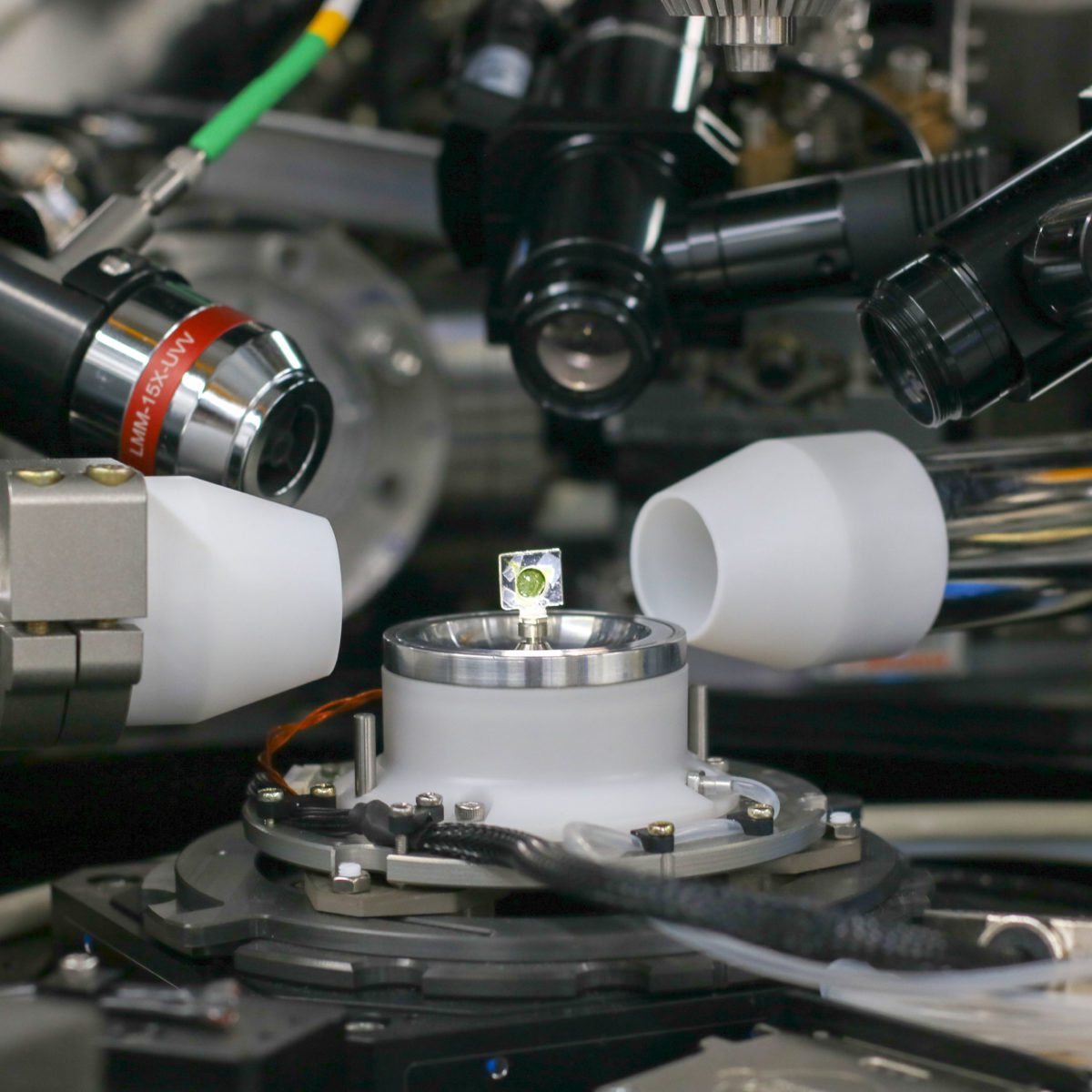
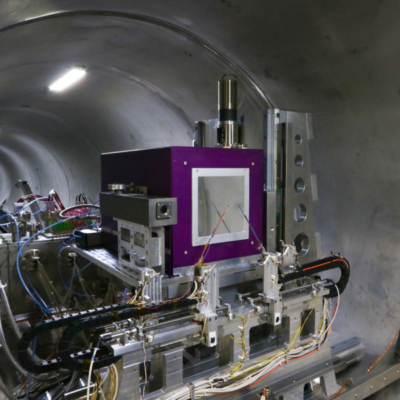
The station, shown in Figure 1, has detectors for the simultaneous techniques such as X-ray fluorescence, X-ray diffraction, X-ray excited optical luminescence and X-ray ptychography, as well as a spectrometer for luminescence and optical microscopes for sample alignment. The station also has a cryogenic assembly, essential for scientific commissioning on samples sensitive to radiation damage. Due to the limitations of the undulator currently installed on the beamline, commissioning of the station is being done in the excitation energy range above 6.4 keV to 14 keV. The lowest achievable fluorescence emission energy now is around 2.0 keV.
Figure 1. TARUMÃ station set up for the first CARNAÚBA experiments in December 2021.
Combination of X-ray fluorescence and ptychography – spatial resolution
Figure 2 shows the results of two types of measurements: the first, on the right, is obtained by measuring the gold (Au) emission line with the fluorescence detectors; the second is the result of a measurement in transmission mode using the PiMega detector and subsequent reconstruction of the image using algorithms to obtain the phase of the wave. This technique takes advantage of the spatial coherence of the beam and is known as ptychography.
While the resolution of the fluorescence image is limited by the size of the beam at the focus, in this case about 120 nm x 250 nm, the image resolution in X-ray ptychography can be increased by exploiting the coherence effect of Sirius’ synchrotron light beam. The image on the left shows a resolution of the order of 20 nm obtained by ptychographic reconstruction of the phase of the wave propagating to the 2D PiMega detector.
These images with this spatial resolution represent an important step in the commissioning of the experimental station, certifying: (i) the simultaneous functionality of the fluorescence and image detectors; (ii) image reconstruction algorithms by X-ray ptychography; (iii) the quality of focus and of the spatial resolution of the beam; (iv) the fast flyscan procedure; and (v) stability in the positioning between the X-ray nanoprobe and the sample, which must be at the level of resolution obtained by ptychography.
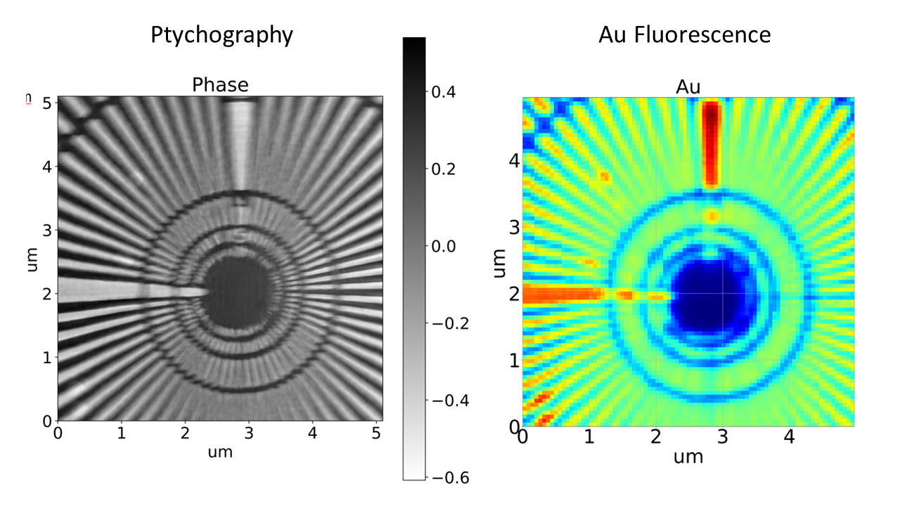
Figure 2. Scanning image of the star pattern (ANT pattern) on the CARNAÚBA beamline, simultaneously measuring fluorescence and ptychography. Image obtained in “pink beam” mode with a beam focused at 120 x 250 nm2 (VxH) from Au fluorescence (right) and by ptychographic reconstruction (phase) with a pixel of 18 nm and an estimated resolution of 20 nm by FRC.
Two-dimensional X-ray fluorescence mapping in soils, roots and seeds
At this stage of commissioning, although the beam is still slightly larger than nominally expected, it is already possible to obtain chemical mapping of complex samples, such as the example of roots in soil shown in Figure 3. It is possible to observe in optical microscopy, on the left, a root containing grains of soil around it in some regions. Using this image with a larger field of view as a reference, the region indicated by the yellow square on the X-ray nanoprobe was centered and an elemental mapping was performed by X-ray fluorescence, with a field of view of 100 µm x 100 µm and 1 µm step. This result is shown in the central image. The right image presents the fluorescence spectra in three distinct regions in the sample, and the lower part presents the different correlations between the most relevant chemical elements.
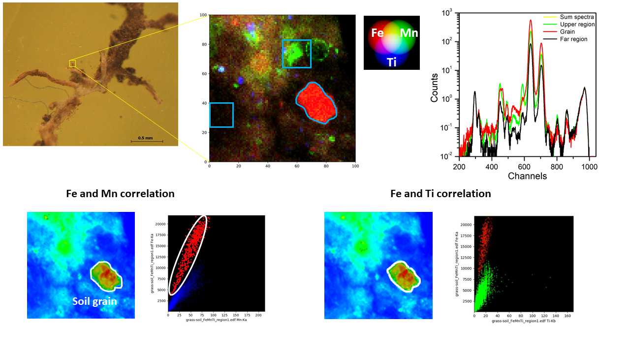
Figure 3. Fluorescence image of a root and soil sample obtained from the CARNAÚBA beamline, with position correlations between different chemical elements.
The instrumental conditions of the beamline already allow carrying out two-dimensional (2D) scanning experiments and obtaining images by attenuation and fluorescence contrast, as well as choosing regions of interest in the samples for X-ray absorption spectroscopy measurements. An example of 2D mapping by fluorescence, observing the distribution of the micronutrients zinc (Zn) and selenium (Se) in a Brazil nut is shown in Figure 4. On the top-left, the optical image is presented in a region of a few square millimeters and below the fluorescence image of 80 µm x 80 µm with 800 nm step. On the top- right, the integrated fluorescence spectrum is shown, where both Zn and Se are highlighted. The chemical speciation of the element Selenium, for the identification of its oxidation state, was performed by energy scanning of the monochromator, keeping the beam position in a fixed position of the sample. The submicrometer resolution of this study was performed with the beam focused at 120 nm x 500 nm. The presence of organic selenium in the -2 oxidation state is clearly observed. This determination was made by comparison with known standard samples.
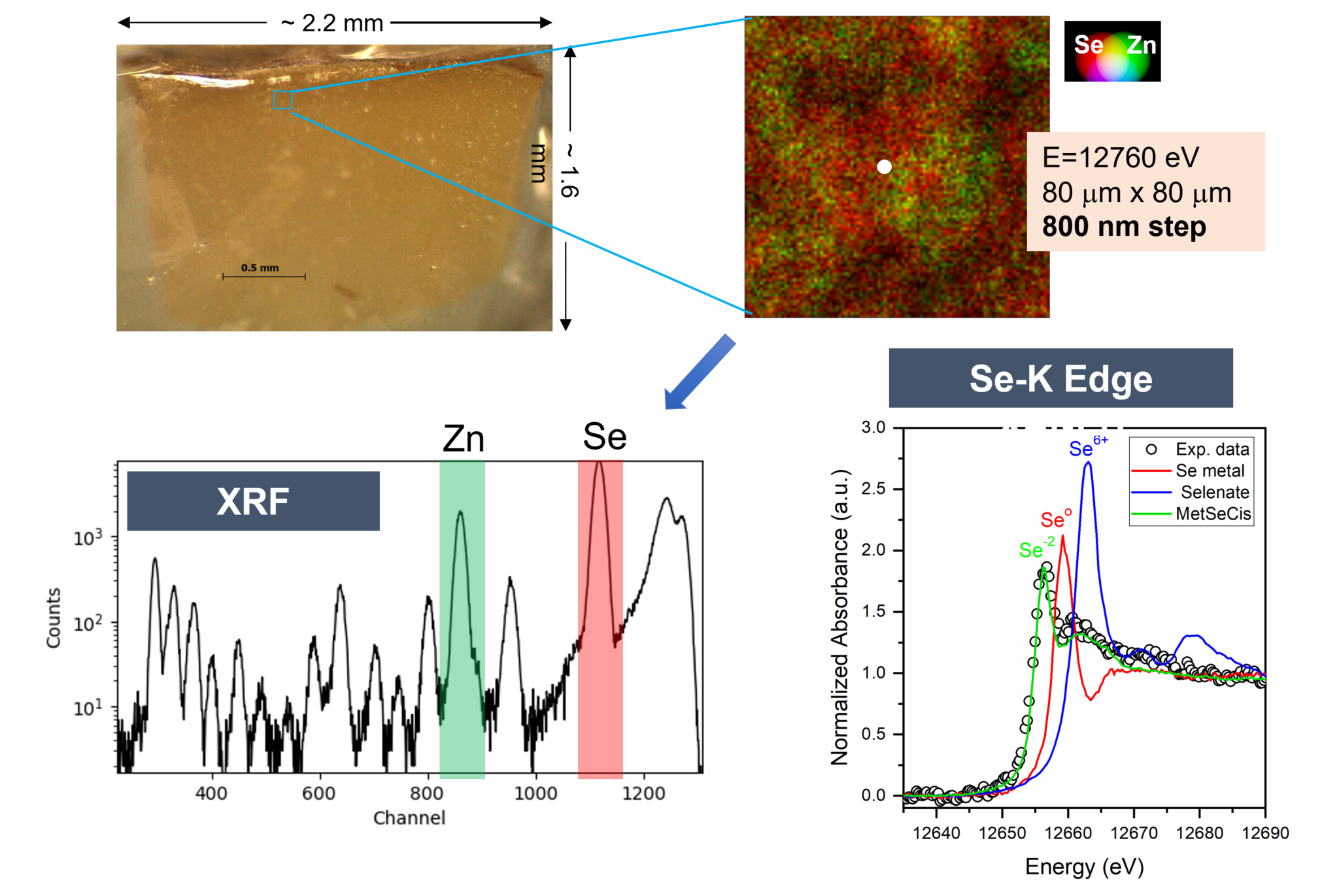
Figure 4. Fluorescence and chemical speciation image of a Brazil nut sample obtained in the CARNAÚBA beamline. The 2D fluorescence image was made by observing the micronutrients Zn and Se, their distributions and correlations. Chemical speciation was done at the X-ray absorption edge of the element Se, indicating that its most likely chemical form is organic.
Tomography with simultaneous attenuation and fluorescence contrast
The first experiment for commissioning the tomography technique on the CARNAÚBA beamline was carried out on a deep diamond sample. Deep diamonds are like time capsules that bring the history of their formation to the Earth’s surface. They trap mineral inclusions, of varied composition, which, through their chemical composition and structure, provide information about many geochemical and geophysical processes that could not be studied in any other way.
The diamonds studied in the CARNAÚBA beamline, which came from a collaboration with the Federal University of Goiás and the University of Brasília, originate from the Juína region in Mato Grosso do Sul, known for the abundance of these objects. These deep diamond samples are estimated to originate from between 400 and 600 km deep in the Earth’s crust. Typical dimensions of the diamonds studied here are on the order of a few millimeters.
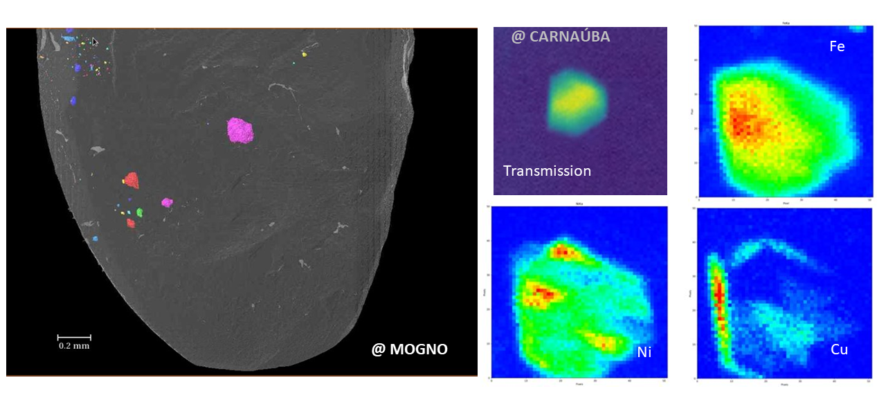
Figure 5. Combining the MOGNO and CARNAÚBA beamlines for the study of mineral inclusions in deep diamonds. Left: Tomography performed on the MOGNO beamline, showing the segmentation result of some solid mineral inclusions. Right: 2D image of transmission and X-ray fluorescence mapping, showing the chemical composition and one of the inclusions.
The diamond sample was previously studied by high-energy X-ray tomography in the MOGNO beamline to identify inclusions and their spatial distribution (Figure 5, left). Once the inclusions, or regions of abundance of inclusions, have been identified, it is possible to use the TARUMÃ station to carry out transmission and X-ray fluorescence mapping (Figure 5, right) and identify in more detail the chemical composition and morphology of these inclusions. In the projection of the tomographic image collected in the MOGNO beamline, an inclusion within the diamond was selected with dimensions compatible with the field of view available in the CARNAÚBA beamline, that is, in this case between 20 and 40 µm. Figure 5, on the right, presents the images of this solid inclusion of approximately 20 µm by attenuation (transmission) contrast, where the total field of view is 50 µm x 50 µm, with a step of 500 nm. The fluorescence contrast shows that this metallic microcrystal is basically formed by atoms of iron (Fe), nickel (Ni) and copper (Cu). Something very unexpected about this mixture of chemical elements is that the Fe-Ni microcrystal is covered by a very inhomogeneous copper layer.

Figure 6 – Computed tomography in the x-ray transmission mode at the TARUMÃ station of the CARNAÚBA beamline.

Figure 7 – Computed tomography in the x-ray fluorescence mode, showing the Cu coverage in relation to the microcrystal, at the TARUMÃ station of the CARNAÚBA beamline.
Figures 6 and 7 show the transmitted and fluorescence tomographic images performed by acquiring 150 projections equivalent to the one shown in Figure 5 and subsequent reconstruction by computed tomography. The image clearly demonstrates that, in fact, the Fe-Ni microcrystal (Fig.6) is covered by a Cu layer and that this covering clearly depends on the face of the crystal (Fig.7). This microcrystal image was the first tomographic image reconstructed in the CARNAÚBA beamline.
Cryogenic system for the study of samples sensitive to radiation damage
The CARNAÚBA beamline allows the study of a wide variety of materials, especially materials with a hybrid composition – organic and inorganic – and biological. These materials are generally sensitive to the radiation doses commonly required for studies, which results in chemical and structural damage and, consequently, changes in their properties. Therefore, these experiments are only possible for cryogenic cooled samples and in inert atmospheres to mitigate the effects of radiation.
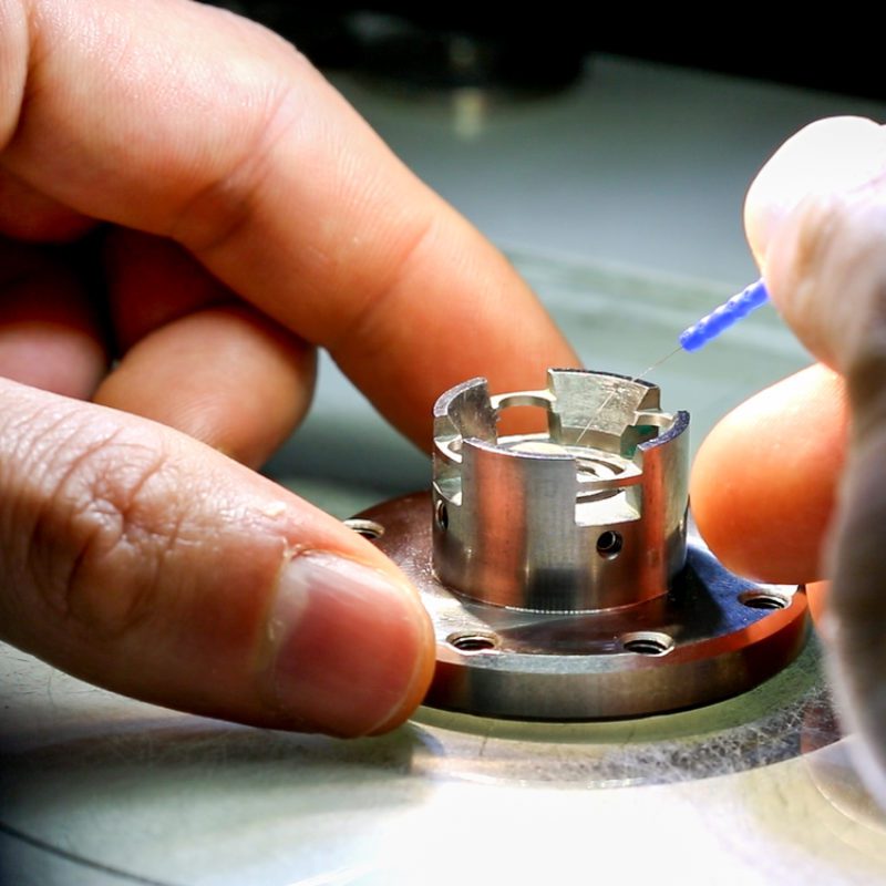
At TARUMÃ, to suit the highly occupied space around the sample and its positioning architecture, an open atmosphere cryogenic system was developed based on a commercial Cryojet, a gas recovery system and a custom sample holder. Following a predictive engineering approach, the system can condition the sample to 150 K, preventing freezing and condensation on nearby instrumentation and preserving the station’s nano-level positioning requirements.
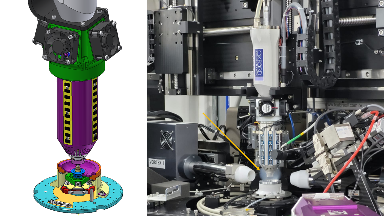
Figure 8. Schematic (left) and photograph (right) of the cryogenics system of the TARUMÃ station on the CARNAÚBA beamline (Lena et al., MEDSI 2021, doi:10.18429/JACoW-MEDSI2020-WEPC02). The arrow indicates the region of cryogenic N2 gas around the sample.
The system was installed and commissioned at the experimental station, indicating that the disturbances produced are within the design parameters for the beamline, being below 4 nm horizontally and 7 nm vertically. The first tests with sensitive samples of hybrid organic-inorganic perovskites clearly demonstrated the need for cooling the samples to increase their resilience to radiation.
Adhesive sealed microfluidic device for in-situ analytical measurements
Microfluidic devices operate with small amounts of the sample through micrometric channels. As main characteristics, these devices have a reduced size, low weight, and high performance, ideal for use in nanoprobes. These characteristics have attracted a lot of attention from the synchrotron community interested in compatible instrumentation for in-situ, operando and in-vivo experiments.
In this work, recently published in Scientific Reports, the team from the CARNAÚBA beamline, in collaboration with LNNano, presented the microfabrication and characterization of a multifunctional microfluidic device well suited to combine X-ray analytical techniques. The device consists of microchannels in glass, where three gold electrodes are deposited in the channels, to serve in-situ electrochemistry, analysis, or standard electrical measurements, sealed by a polyester film. The device demonstrated excellent chemical resistance to organic solvents, good resistance to pressure, and its efficiency in the presence of biological samples (proteins) is remarkable. It was characterized with synchrotron light through 2D X-ray fluorescence mapping of both its basic components, such as the gold electrode (Au) and the chromium adhesion film (Cr), as well as the result of an electrochemical reaction involving silver (Ag) and chlorine (Cl) and the nucleation of Ag nanoparticles at the Au electrode.
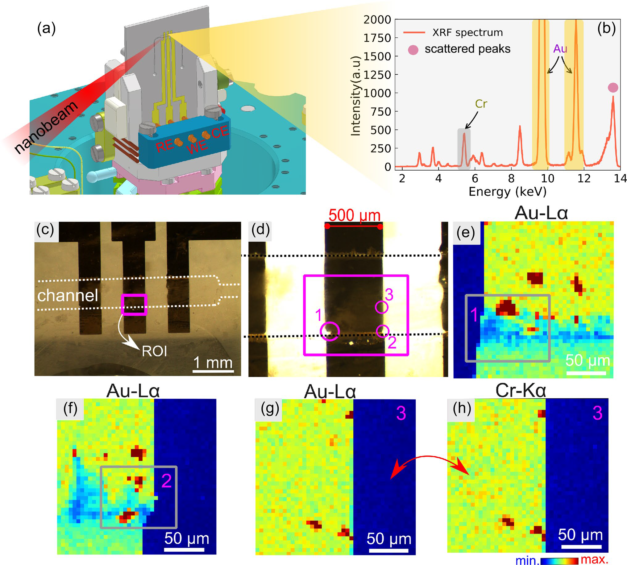
Figure 9. Nanofluorescence experiments on the microfluidic device. (a) 3D schematic of the microfluidic sample holder. (b) Fluorescence spectrum collected at the central electrode. (c-d) Optical micrograph of the channel and electrodes highlighting the ROI (magenta rectangle) scanned by a nanobeam of about 600 nm x 600 nm. (e-f) Au-La fluorescence maps (9713 eV) over circle 1 and 2 at the edge of the electrode. (g-h) Au (La) and Cr-Ka (5.4 keV) maps obtained in circle 3. The experiments were carried out in the channels filled with water. Nano-XRF maps have a pixel size of 5 µm and a total acquisition time of 65 seconds, with 22.5 ms per pixel or step (I. Neckel et al., Scientific Reports, 2021).
Electrochemical cell for the in-situ study of materials for (electro)-catalysis and batteries
Materials for (electro-)catalysis and batteries play a crucial role in the present and future of a sustainable and clean economy. Unraveling its structure and understanding its functioning, especially when in the form of nanomaterials, is a basic requirement to obtain increasingly adapted and efficient materials.
To this end, the CARNAÚBA beamline team is developing a cell for in-situ electrochemistry studies of the synthesis of these materials. This cell is compatible with X-ray absorption experiments in fluorescence mode and diffraction experiments, whether or not exploring probe coherence. One technique that will be widely applied to nanocrystal systems is the Bragg CDI technique, which provides information about the displacement field of atoms in the crystal structure, which is directly related to voltage.
The cell, shown in Figure 10, has already been assembled and certified from an electrochemical point of view compared to conventional laboratory cells and will soon be used in in-situ experiments of electrochemical nanocrystal growth.
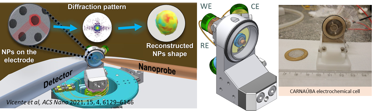
Figure 10. Schematic of the electrochemical cell mounted on the CARNAÚBA beamline showing the measurement geometry and the strategy to obtain, from coherent Bragg diffraction, the displacement field of atoms in nanoparticles (Vicente, Neckel, Fernandez et al., ACS Nano, 2021). In the center is shown the design of the electrochemical cell and on the right its construction and assembly (Wilendorf et al., MEDSI 2021, doi:10.18429/JACoW-MEDSI2020-WEPC03).
First 3D imaging experiments at Sirius were performed using ptychographic nanotomography
This call for proposals intends to supply beamtime for the users during the beamline commissioning period